ARG63217
anti-RGS1 / 1R20 antibody
anti-RGS1 / 1R20 antibody for IHC-Formalin-fixed paraffin-embedded sections,Western blot and Human
Signaling Transduction antibody
Overview
| Product Description | Goat Polyclonal antibody recognizes RGS1 / 1R20 |
|---|---|
| Tested Reactivity | Hu |
| Predict Reactivity | Ms, Rat, Cow, Dog |
| Tested Application | IHC-P, WB |
| Host | Goat |
| Clonality | Polyclonal |
| Isotype | IgG |
| Target Name | RGS1 / 1R20 |
| Antigen Species | Human |
| Immunogen | C-NLLNDLQANSLK |
| Conjugation | Un-conjugated |
| Alternate Names | Regulator of G-protein signaling 1; 1R20; IER1; IR20; B-cell activation protein BL34; RGS1; BL34; Early response protein 1R20; HEL-S-87 |
Application Instructions
| Application Suggestion |
|
||||||
|---|---|---|---|---|---|---|---|
| Application Note | WB: Recommend incubate at RT for 1h. IHC-P: Antigen Retrieval: Steam tissue section in Citrate buffer (pH 6.0). * The dilutions indicate recommended starting dilutions and the optimal dilutions or concentrations should be determined by the scientist. |
Properties
| Form | Liquid |
|---|---|
| Purification | Purified from goat serum by ammonium sulphate precipitation followed by antigen affinity chromatography using the immunizing peptide. |
| Buffer | Tris saline (pH 7.3), 0.02% Sodium azide and 0.5% BSA |
| Preservative | 0.02% Sodium azide |
| Stabilizer | 0.5% BSA |
| Concentration | 0.5 mg/ml |
| Storage Instruction | For continuous use, store undiluted antibody at 2-8°C for up to a week. For long-term storage, aliquot and store at -20°C or below. Storage in frost free freezers is not recommended. Avoid repeated freeze/thaw cycles. Suggest spin the vial prior to opening. The antibody solution should be gently mixed before use. |
| Note | For laboratory research only, not for drug, diagnostic or other use. |
Bioinformation
| Database Links |
Swiss-port # Q08116 Human Regulator of G-protein signaling 1 |
|---|---|
| Background | This gene encodes a member of the regulator of G-protein signalling family. This protein is located on the cytosolic side of the plasma membrane and contains a conserved, 120 amino acid motif called the RGS domain. The protein attenuates the signalling activity of G-proteins by binding to activated, GTP-bound G alpha subunits and acting as a GTPase activating protein (GAP), increasing the rate of conversion of the GTP to GDP. This hydrolysis allows the G alpha subunits to bind G beta/gamma subunit heterodimers, forming inactive G-protein heterotrimers, thereby terminating the signal. [provided by RefSeq, Jul 2008] |
| Research Area | Signaling Transduction antibody |
| Calculated MW | 24 kDa |
Images (3) Click the Picture to Zoom In
-
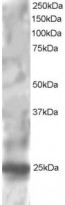
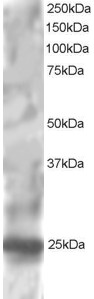
ARG63217 anti-RGS1 / 1R20 antibody WB image
Western Blot: HepG2 lysate (RIPA buffer, 30µg total protein per lane) stained with ARG63217 anti-RGS1 / 1R20 antibody at 1 µg/ml dilution.
-

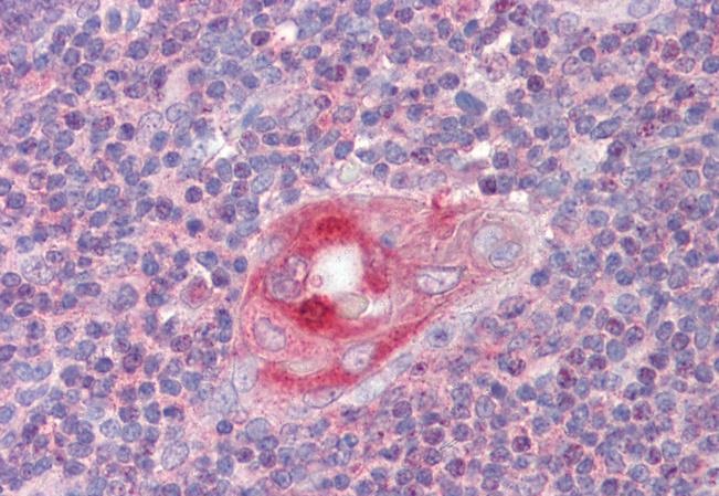
ARG63217 anti-RGS1 / 1R20 antibody IHC-P image
Immunohistochemistry: Paraffin-embedded Human thymus tissue. Antigen Retrieval: Steam tissue section in Citrate buffer (pH 6.0). The tissue section was stained with ARG63217 anti-RGS1 / 1R20 antibody at 2.5 µg/ml dilution followed by AP-staining.
-
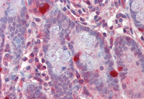
ARG63217 anti-RGS1 / 1R20 antibody IHC-P image
Immunohistochemistry: Paraffin-embedded Human small intestine tissue. Antigen Retrieval: Steam tissue section in Citrate buffer (pH 6.0). The tissue section was stained with ARG63217 anti-RGS1 / 1R20 antibody at 2.5 µg/ml dilution followed by AP-staining.