ARG43136
anti-RBPJK antibody
anti-RBPJK antibody for Flow cytometry,ICC/IF,Western blot and Human,Mouse,Rat
Overview
| Product Description | Rabbit Polyclonal antibody recognizes RBPJK |
|---|---|
| Tested Reactivity | Hu, Ms, Rat |
| Tested Application | FACS, ICC/IF, WB |
| Host | Rabbit |
| Clonality | Polyclonal |
| Isotype | IgG |
| Target Name | RBPJK |
| Antigen Species | Human |
| Immunogen | Recombinant protein corresponding to K41-Q467 of Human RBPJK. |
| Conjugation | Un-conjugated |
| Alternate Names | CBF1; csl; IGKJRB1; RBPJK; IGKJRB; RBP-J kappa; Recombining binding protein suppressor of hairless; SUH; J kappa-recombination signal-binding protein; AOS3; RBP-J; RBP-JK; KBF2; CBF-1; RBPSUH; Renal carcinoma antigen NY-REN-30 |
Application Instructions
| Application Suggestion |
|
||||||||
|---|---|---|---|---|---|---|---|---|---|
| Application Note | * The dilutions indicate recommended starting dilutions and the optimal dilutions or concentrations should be determined by the scientist. | ||||||||
| Observed Size | ~ 58 kDa |
Properties
| Form | Liquid |
|---|---|
| Purification | Immunogen affinity purified. |
| Buffer | 0.2% Na2HPO4, 0.9% NaCl, 0.05% Sodium azide and 4% Trehalose. |
| Preservative | 0.05% Sodium azide |
| Stabilizer | 4% Trehalose |
| Concentration | 0.5 mg/ml |
| Storage Instruction | For continuous use, store undiluted antibody at 2-8°C for up to a week. For long-term storage, aliquot and store at -20°C or below. Storage in frost free freezers is not recommended. Avoid repeated freeze/thaw cycles. Suggest spin the vial prior to opening. The antibody solution should be gently mixed before use. |
| Note | For laboratory research only, not for drug, diagnostic or other use. |
Bioinformation
| Database Links |
Swiss-port # P31266 Mouse Recombining binding protein suppressor of hairless Swiss-port # Q06330 Human Recombining binding protein suppressor of hairless |
|---|---|
| Gene Symbol | RBPJ |
| Gene Full Name | recombination signal binding protein for immunoglobulin kappa J region |
| Background | The protein encoded by this gene is a transcriptional regulator important in the Notch signaling pathway. The encoded protein acts as a repressor when not bound to Notch proteins and an activator when bound to Notch proteins. It is thought to function by recruiting chromatin remodeling complexes containing histone deacetylase or histone acetylase proteins to Notch signaling pathway genes. Several transcript variants encoding different isoforms have been found for this gene, and several pseudogenes of this gene exist on chromosome 9. [provided by RefSeq, Oct 2013] |
| Function | Transcriptional regulator that plays a central role in Notch signaling, a signaling pathway involved in cell-cell communication that regulates a broad spectrum of cell-fate determinations. Acts as a transcriptional repressor when it is not associated with Notch proteins. When associated with some NICD product of Notch proteins (Notch intracellular domain), it acts as a transcriptional activator that activates transcription of Notch target genes. Probably represses or activates transcription via the recruitment of chromatin remodeling complexes containing histone deacetylase or histone acetylase proteins, respectively. Specifically binds to the immunoglobulin kappa-type J segment recombination signal sequence. Binds specifically to methylated DNA (PubMed:21991380). Binds to the oxygen responsive element of COX4I2 and activates its transcription under hypoxia conditions (4% oxygen) (PubMed:23303788). Negatively regulates the phagocyte oxidative burst in response to bacterial infection by repressing transcription of NADPH oxidase subunits (By similarity). [UniProt] |
| Cellular Localization | Nucleus. Cytoplasm. Note=Mainly nuclear, upon interaction with RITA/C12orf52, translocates to the cytoplasm, down-regulating the Notch signaling pathway. [UniProt] |
| Calculated MW | 56 kDa |
Images (3) Click the Picture to Zoom In
-
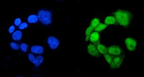
ARG43136 anti-RBPJK antibody ICC/IF image
Immunofluorescence: A431 cells were blocked with 10% goat serum and then stained with ARG43136 anti-RBPJK antibody (green) at 5 µg/ml dilution, overnight at 4°C. DAPI (blue) for nuclear staining.
-
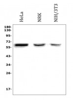
ARG43136 anti-RBPJK antibody WB image
Western blot: 50 µg of sample under reducing conditions. HeLa, NRK and NIH/3T3 whole cell lysates stained with ARG43136 anti-RBPJK antibody at 0.5 µg/ml dilution, overnight at 4°C.
-
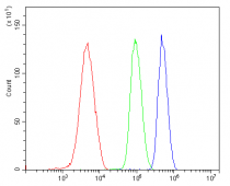
ARG43136 anti-RBPJK antibody FACS image
Flow Cytometry: HL-60 cells were blocked with 10% normal goat serum and then stained with ARG43136 anti-RBPJK antibody (blue) at 1 µg/10^6 cells for 30 min at 20°C, followed by incubation with DyLight®488 labelled secondary antibody. Isotype control antibody (green) was rabbit IgG (1 µg/10^6 cells) used under the same conditions. Unlabelled sample (red) was also used as a control.


