ARG59015
anti-RALBP1 antibody
anti-RALBP1 antibody for Flow cytometry,ICC/IF,IHC-Formalin-fixed paraffin-embedded sections,Western blot and Human,Mouse,Rat
Overview
| Product Description | Rabbit Polyclonal antibody recognizes RALBP1 |
|---|---|
| Tested Reactivity | Hu, Ms, Rat |
| Tested Application | FACS, ICC/IF, IHC-P, WB |
| Host | Rabbit |
| Clonality | Polyclonal |
| Isotype | IgG |
| Target Name | RALBP1 |
| Antigen Species | Human |
| Immunogen | Recombinant protein corresponding to K239-Q506 of Human RALBP1. |
| Conjugation | Un-conjugated |
| Alternate Names | RalA-binding protein 1; RalBP1; Dinitrophenyl S-glutathione ATPase; DNP-SG ATPase; RIP1; RLIP76; Ral-interacting protein 1; 76 kDa Ral-interacting protein; RLIP1 |
Application Instructions
| Application Suggestion |
|
||||||||||
|---|---|---|---|---|---|---|---|---|---|---|---|
| Application Note | IHC-P: Antigen Retrieval: Heat mediation was performed in Citrate buffer (pH 6.0) for 20 min. * The dilutions indicate recommended starting dilutions and the optimal dilutions or concentrations should be determined by the scientist. |
Properties
| Form | Liquid |
|---|---|
| Purification | Affinity purification with immunogen. |
| Buffer | 0.2% Na2HPO4, 0.9% NaCl, 0.05% Sodium azide and 4% Trehalose. |
| Preservative | 0.05% Sodium azide |
| Stabilizer | 4% Trehalose |
| Concentration | 0.5 mg/ml |
| Storage Instruction | For continuous use, store undiluted antibody at 2-8°C for up to a week. For long-term storage, aliquot and store at -20°C or below. Storage in frost free freezers is not recommended. Avoid repeated freeze/thaw cycles. Suggest spin the vial prior to opening. The antibody solution should be gently mixed before use. |
| Note | For laboratory research only, not for drug, diagnostic or other use. |
Bioinformation
| Database Links | |
|---|---|
| Gene Symbol | RALBP1 |
| Gene Full Name | ralA binding protein 1 |
| Background | RALBP1 plays a role in receptor-mediated endocytosis and is a downstream effector of the small GTP-binding protein RAL (see RALA; MIM 179550). Small G proteins, such as RAL, have GDP-bound inactive and GTP-bound active forms, which shift from the inactive to the active state through the action of RALGDS (MIM 601619), which in turn is activated by RAS (see HRAS; MIM 190020) (summary by Feig, 2003 [PubMed 12888294]).[supplied by OMIM, Nov 2010] |
| Function | Can activate specifically hydrolysis of GTP bound to RAC1 and CDC42, but not RALA. Mediates ATP-dependent transport of S-(2,4-dinitrophenyl)-glutathione (DNP-SG) and doxorubicin (DOX) and is the major ATP-dependent transporter of glutathione conjugates of electrophiles (GS-E) and DOX in erythrocytes. Can catalyze transport of glutathione conjugates and xenobiotics, and may contribute to the multidrug resistance phenomenon. Serves as a scaffold protein that brings together proteins forming an endocytotic complex during interphase and also with CDK1 to switch off endocytosis, One of its substrates would be EPN1/Epsin. [UniProt] |
| Cellular Localization | Membrane. [UniProt] |
| Calculated MW | 76 kDa |
Images (7) Click the Picture to Zoom In
-

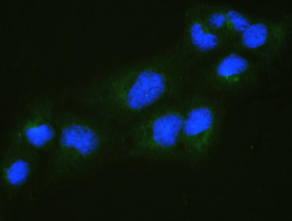
ARG59015 anti-RALBP1 antibody ICC/IF image
Immunofluorescence: U2OS cells were blocked with 10% goat serum and then stained with ARG59015 anti-RALBP1 antibody (green) at 2 µg/ml dilution, overnight at 4°C. DAPI (blue) for nuclear staining.
-
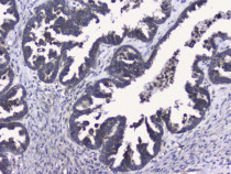
ARG59015 anti-RALBP1 antibody IHC-P image
Immunohistochemistry: Paraffin-embedded Human ovary cancer tissue. Antigen Retrieval: Heat mediation was performed in Citrate buffer (pH 6.0) for 20 min. The tissue section was blocked with 10% goat serum. The tissue section was then stained with ARG59015 anti-RALBP1 antibody at 1 µg/ml dilution, overnight at 4°C.
-

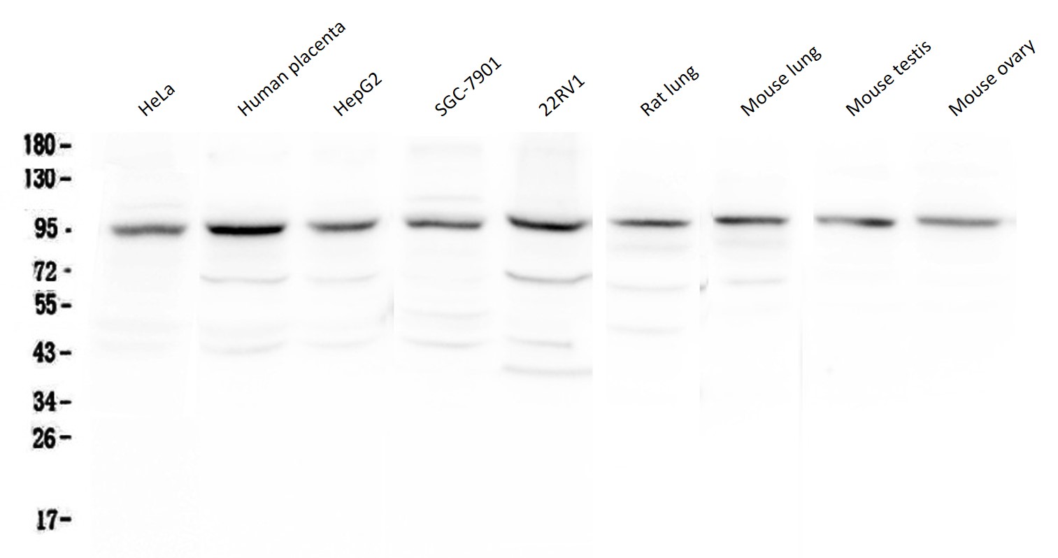
ARG59015 anti-RALBP1 antibody WB image
Western blot: 50 µg of samples under reducing conditions. HeLa, Human placenta, HepG2, SGC-7901, 22RV1, Rat lung, Mouse lung, Mouse testis and Mouse ovary lysates stained with ARG59015 anti-RALBP1 antibody at 0.5 µg/ml, overnight at 4°C.
-
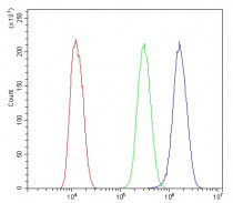
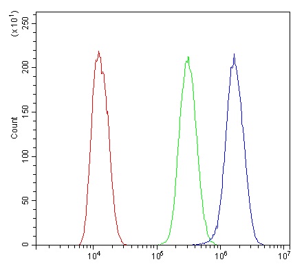
ARG59015 anti-RALBP1 antibody FACS image
Flow Cytometry: U2OS cells were blocked with 10% normal goat serum and then stained with ARG59015 anti-RALBP1 antibody (blue) at 1 µg/10^6 cells for 30 min at 20°C, followed by incubation with DyLight®488 labelled secondary antibody. Isotype control antibody (green) was rabbit IgG (1 µg/10^6 cells) used under the same conditions. Unlabelled sample (red) was also used as a control.
-
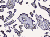
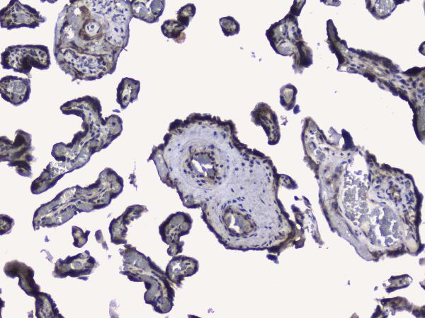
ARG59015 anti-RALBP1 antibody IHC-P image
Immunohistochemistry: Paraffin-embedded Human placenta tissue. Antigen Retrieval: Heat mediation was performed in Citrate buffer (pH 6.0) for 20 min. The tissue section was blocked with 10% goat serum. The tissue section was then stained with ARG59015 anti-RALBP1 antibody at 1 µg/ml dilution, overnight at 4°C.
-
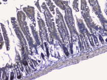
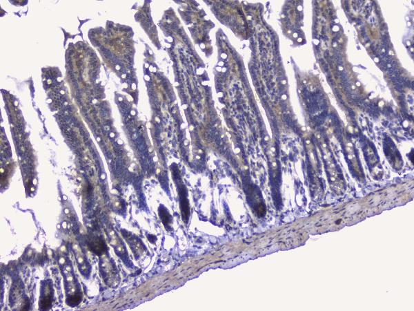
ARG59015 anti-RALBP1 antibody IHC-P image
Immunohistochemistry: Paraffin-embedded Mouse small intestine tissue. Antigen Retrieval: Heat mediation was performed in Citrate buffer (pH 6.0) for 20 min. The tissue section was blocked with 10% goat serum. The tissue section was then stained with ARG59015 anti-RALBP1 antibody at 1 µg/ml dilution, overnight at 4°C.
-
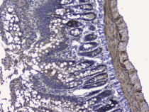
ARG59015 anti-RALBP1 antibody IHC-P image
Immunohistochemistry: Paraffin-embedded Rat small intestine tissue. Antigen Retrieval: Heat mediation was performed in Citrate buffer (pH 6.0) for 20 min. The tissue section was blocked with 10% goat serum. The tissue section was then stained with ARG59015 anti-RALBP1 antibody at 1 µg/ml dilution, overnight at 4°C.