ARG63188
anti-RACGAP1 / MgcRacGAP antibody
anti-RACGAP1 / MgcRacGAP antibody for Flow cytometry,ICC/IF,IHC-Formalin-fixed paraffin-embedded sections,Western blot and Human
Cell Biology and Cellular Response antibody; Signaling Transduction antibody
Overview
| Product Description | Goat Polyclonal antibody recognizes RACGAP1 / MgcRacGAP |
|---|---|
| Tested Reactivity | Hu |
| Predict Reactivity | Cow, Dog |
| Tested Application | FACS, ICC/IF, IHC-P, WB |
| Specificity | Reported variants represent identical protein: NP_037409.2, NP_001119576.1 and NP_001119575.1. |
| Host | Goat |
| Clonality | Polyclonal |
| Isotype | IgG |
| Target Name | RACGAP1 / MgcRacGAP |
| Antigen Species | Human |
| Immunogen | C-GRQGNFFASPMLK |
| Conjugation | Un-conjugated |
| Alternate Names | CYK4; Rac GTPase-activating protein 1; MgcRacGAP; Protein CYK4 homolog; Male germ cell RacGap; ID-GAP; HsCYK-4 |
Application Instructions
| Application Suggestion |
|
||||||||||
|---|---|---|---|---|---|---|---|---|---|---|---|
| Application Note | WB: Recommend incubate at RT for 1h. IHC-P: Antigen Retrieval: Microwaved tissue section in Tris/EDTA buffer (pH 9.0). * The dilutions indicate recommended starting dilutions and the optimal dilutions or concentrations should be determined by the scientist. |
Properties
| Form | Liquid |
|---|---|
| Purification | Purified from goat serum by antigen affinity chromatography. |
| Buffer | Tris saline (pH 7.3), 0.02% Sodium azide and 0.5% BSA. |
| Preservative | 0.02% Sodium azide |
| Stabilizer | 0.5% BSA |
| Concentration | 0.5 mg/ml |
| Storage Instruction | For continuous use, store undiluted antibody at 2-8°C for up to a week. For long-term storage, aliquot and store at -20°C or below. Storage in frost free freezers is not recommended. Avoid repeated freeze/thaw cycles. Suggest spin the vial prior to opening. The antibody solution should be gently mixed before use. |
| Note | For laboratory research only, not for drug, diagnostic or other use. |
Bioinformation
| Database Links | |
|---|---|
| Background | The protein encoded by this gene belongs to the GTPase-activating protein (GAP) family. GAPs bind activated forms of Rho GTPases and stimulate GTP hydrolysis. Through this catalytic function, GAPs negatively regulate Rho-mediated signals. This protein plays a regulatory role in initiation of cytokinesis, controlling cell growth and differentiation of hematopoietic cells, regulating spermatogenesis, and in neuronal proliferation. Alternatively spliced transcript variants have been found for this gene. [provided by RefSeq, Sep 2011] |
| Research Area | Cell Biology and Cellular Response antibody; Signaling Transduction antibody |
| Calculated MW | 71 kDa |
| PTM | Phosphorylated at multiple sites in the midbody during cytokinesis. Phosphorylation by AURKB on Ser-387 at the midbody is, at least in part, responsible for exerting its latent GAP activity towards RhoA. Phosphorylation on multiple serine residues by PLK1 enhances its association with ECT2 and is critical for cleavage furrow formation. |
Images (6) Click the Picture to Zoom In
-
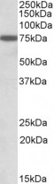
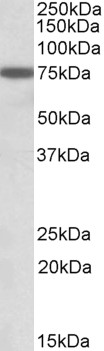
ARG63188 anti-RACGAP1 / MgcRacGAP antibody WB image
Western Blot: K562 cell lysate (35 µg protein in RIPA buffer). stained with ARG63188 anti-RACGAP1 / MgcRacGAP antibody at 1 µg/ml dilution.
-

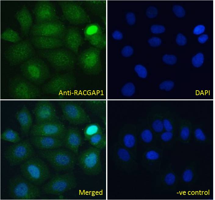
ARG63188 anti-RACGAP1 / MgcRacGAP antibody ICC/IF image
Immunofluorescence: Paraformaldehyde fixed MCF7 cells permeabilized with 0.15% Triton. Cells were stained with ARG63188 anti-RACGAP1 / MgcRacGAP antibody (green) at 10 µg/ml dilution for 1 hour. DAPI (blue) for nuclear staining. Negative control: Unimmunized goat IgG (green) at 10 µg/ml dilution.
-

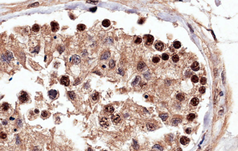
ARG63188 anti-RACGAP1 / MgcRacGAP antibody IHC-P image
Immunohistochemistry: Paraffin-embedded Human testis tissue. Antigen Retrieval: Microwaved tissue section in Tris/EDTA buffer (pH 9.0). The tissue section was stained with ARG63188 anti-RACGAP1 / MgcRacGAP antibody at 4 µg/ml dilution followed by HRP-staining.
-
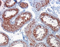
ARG63188 anti-RACGAP1 / MgcRacGAP antibody IHC-P image
Immunohistochemistry: Paraffin-embedded Human testis tissue. (Microwaved antigen retrieval with Tris/EDTA buffer pH9) stained with ARG63188 anti-RACGAP1 / MgcRacGAP antibody at 1 µg/ml dilution followed by HRP-staining.
-
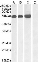
ARG63188 anti-RACGAP1 / MgcRacGAP antibody WB image
Western blot: 35 µg of A431 nucleus (A), Jurkat (B), Jurkat nucleus (C) and Human pancreas (D, negative control) lysates (in RIPA buffer) stained with ARG63188 anti-RACGAP1 / MgcRacGAP antibody at 1 µg/ml dilution and incubated at RT for 1 hour.
-
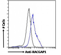
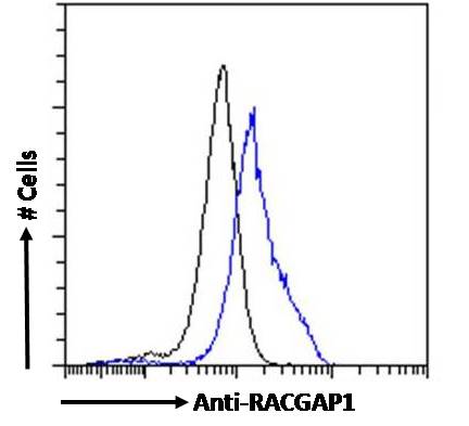
ARG63188 anti-RACGAP1 / MgcRacGAP antibody FACS image
Flow Cytometry: Paraformaldehyde-fixed MCF7 cells permeabilized with 0.5% Triton. Cells were stained with ARG63188 anti-RACGAP1 / MgcRacGAP antibody (blue line) at 10 µg/ml dilution for 1 hour, followed by incubation with Alexa FluorR 488 labelled secondary antibody. IgG control: Unimmunized goat IgG (black line).