ARG43053
anti-RAB3GAP2 antibody
anti-RAB3GAP2 antibody for Flow cytometry,IHC-Formalin-fixed paraffin-embedded sections,Western blot and Human,Mouse,Rat
Overview
| Product Description | Rabbit Polyclonal antibody recognizes RAB3GAP2 |
|---|---|
| Tested Reactivity | Hu, Ms, Rat |
| Tested Application | FACS, IHC-P, WB |
| Host | Rabbit |
| Clonality | Polyclonal |
| Isotype | IgG |
| Target Name | RAB3GAP2 |
| Antigen Species | Human |
| Immunogen | Recombinant protein corresponding to M1-Q115 of Human RAB3GAP2. |
| Conjugation | Un-conjugated |
| Alternate Names | Rab3-GAP p150; p150; RGAP-iso; RAB3-GAP150; Rab3-GAP regulatory subunit; RAB3GAP150; Rab3-GAP150; Rab3 GTPase-activating protein non-catalytic subunit; SPG69; WARBM2; Rab3 GTPase-activating protein 150 kDa subunit |
Application Instructions
| Application Suggestion |
|
||||||||
|---|---|---|---|---|---|---|---|---|---|
| Application Note | IHC-P: Antigen Retrieval: Heat mediation was performed in Citrate buffer (pH 6.0) for 20 min. * The dilutions indicate recommended starting dilutions and the optimal dilutions or concentrations should be determined by the scientist. |
Properties
| Form | Liquid |
|---|---|
| Purification | Affinity purification with immunogen. |
| Buffer | 0.2% Na2HPO4, 0.9% NaCl, 0.05% Sodium azide and 4% Trehalose. |
| Preservative | 0.05% Sodium azide |
| Stabilizer | 4% Trehalose |
| Concentration | 0.5 mg/ml |
| Storage Instruction | For continuous use, store undiluted antibody at 2-8°C for up to a week. For long-term storage, aliquot and store at -20°C or below. Storage in frost free freezers is not recommended. Avoid repeated freeze/thaw cycles. Suggest spin the vial prior to opening. The antibody solution should be gently mixed before use. |
| Note | For laboratory research only, not for drug, diagnostic or other use. |
Bioinformation
| Database Links | |
|---|---|
| Gene Symbol | RAB3GAP2 |
| Gene Full Name | RAB3 GTPase activating protein subunit 2 (non-catalytic) |
| Background | The protein encoded by this gene belongs to the RAB3 protein family, members of which are involved in regulated exocytosis of neurotransmitters and hormones. This protein forms the Rab3 GTPase-activating complex with RAB3GAP1, where it constitutes the regulatory subunit, whereas the latter functions as the catalytic subunit. This gene has the highest level of expression in the brain, consistent with it having a key role in neurodevelopment. Mutations in this gene are associated with Martsolf syndrome. [provided by RefSeq, Oct 2009] |
| Function | Regulatory subunit of a GTPase activating protein that has specificity for Rab3 subfamily (RAB3A, RAB3B, RAB3C and RAB3D). Rab3 proteins are involved in regulated exocytosis of neurotransmitters and hormones. Rab3 GTPase-activating complex specifically converts active Rab3-GTP to the inactive form Rab3-GDP. Required for normal eye and brain development. May participate in neurodevelopmental processes such as proliferation, migration and differentiation before synapse formation, and non-synaptic vesicular release of neurotransmitters. [UniProt] |
| Cellular Localization | Cytoplasm. Note=In neurons, it is enriched in the synaptic soluble fraction. [UniProt] |
| Calculated MW | 156 kDa |
Images (8) Click the Picture to Zoom In
-
ARG43053 anti-RAB3GAP2 antibody IHC-P image
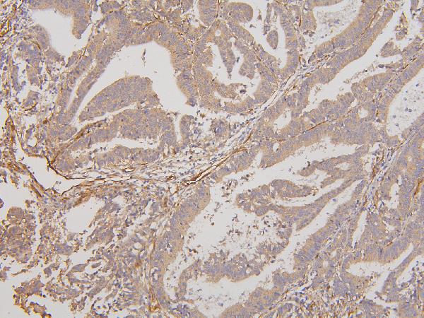
Immunohistochemistry: Paraffin-embedded Human mammary cancer tissue. Antigen Retrieval: Heat mediation was performed in Citrate buffer (pH 6.0) for 20 min. The tissue section was blocked with 10% goat serum. The tissue section was then stained with ARG43053 anti-RAB3GAP2 antibody at 1 µg/ml dilution, overnight at 4°C.
-
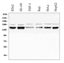
ARG43053 anti-RAB3GAP2 antibody WB image
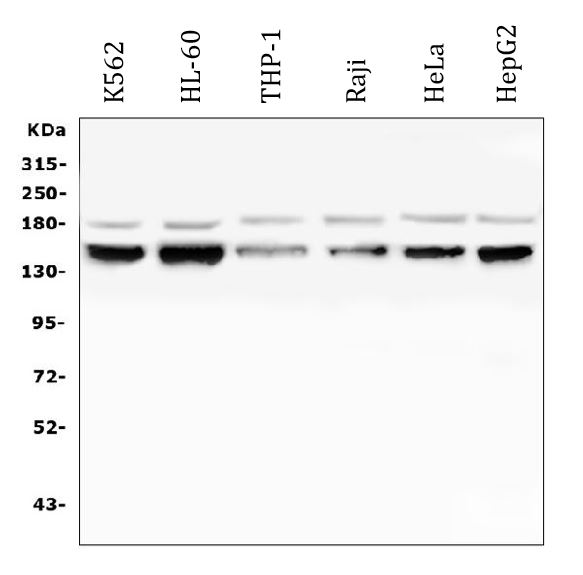
Western blot: 50 µg of sample under reducing conditions. K562, HL-60, THP-1, Raji, HeLa and HepG2 whole cell lysates stained with ARG43053 anti-RAB3GAP2 antibody at 0.5 µg/ml dilution, overnight at 4°C.
-
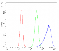
ARG43053 anti-RAB3GAP2 antibody FACS image
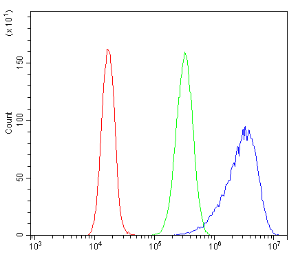
Flow Cytometry: Caco-2 cells were blocked with 10% normal goat serum and then stained with ARG43053 anti-RAB3GAP2 antibody (blue) at 1 µg/10^6 cells for 30 min at 20°C, followed by incubation with DyLight®488 labelled secondary antibody. Isotype control antibody (green) was rabbit IgG (1 µg/10^6 cells) used under the same conditions. Unlabelled sample (red) was also used as a control.
-
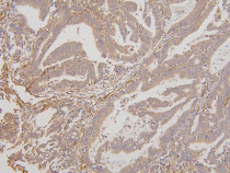
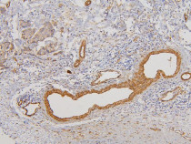
ARG43053 anti-RAB3GAP2 antibody IHC-P image
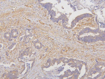
Immunohistochemistry: Paraffin-embedded Human rectal cancer tissue. Antigen Retrieval: Heat mediation was performed in Citrate buffer (pH 6.0) for 20 min. The tissue section was blocked with 10% goat serum. The tissue section was then stained with ARG43053 anti-RAB3GAP2 antibody at 1 µg/ml dilution, overnight at 4°C.
-
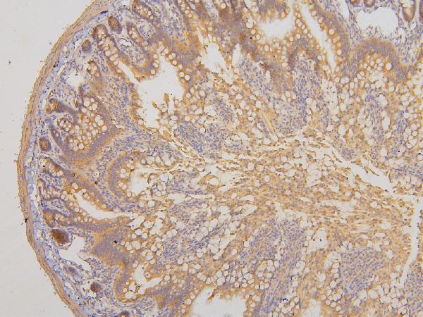
ARG43053 anti-RAB3GAP2 antibody IHC-P image
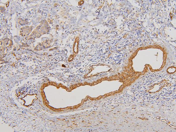
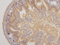
Immunohistochemistry: Paraffin-embedded Mouse intestine tissue. Antigen Retrieval: Heat mediation was performed in Citrate buffer (pH 6.0) for 20 min. The tissue section was blocked with 10% goat serum. The tissue section was then stained with ARG43053 anti-RAB3GAP2 antibody at 1 µg/ml dilution, overnight at 4°C.
-
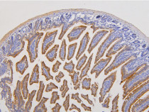
ARG43053 anti-RAB3GAP2 antibody IHC-P image
Immunohistochemistry: Paraffin-embedded Rat intestine tissue. Antigen Retrieval: Heat mediation was performed in Citrate buffer (pH 6.0) for 20 min. The tissue section was blocked with 10% goat serum. The tissue section was then stained with ARG43053 anti-RAB3GAP2 antibody at 1 µg/ml dilution, overnight at 4°C.
-
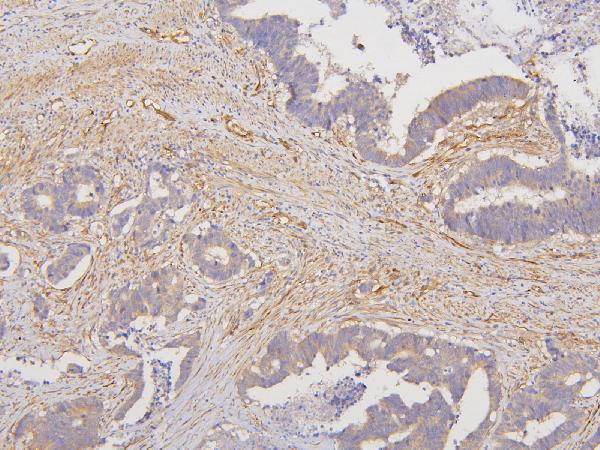
ARG43053 anti-RAB3GAP2 antibody IHC-P image
Immunohistochemistry: Paraffin-embedded Human colon cancer tissue. Antigen Retrieval: Heat mediation was performed in Citrate buffer (pH 6.0) for 20 min. The tissue section was blocked with 10% goat serum. The tissue section was then stained with ARG43053 anti-RAB3GAP2 antibody at 1 µg/ml dilution, overnight at 4°C.
-

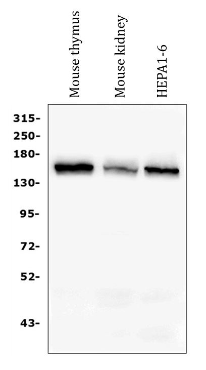
ARG43053 anti-RAB3GAP2 antibody WB image
Western blot: 50 µg of sample under reducing conditions. Mouse thymus, Mouse kidney and HEPA1-6 whole cell lysates stained with ARG43053 anti-RAB3GAP2 antibody at 0.5 µg/ml dilution, overnight at 4°C.