ARG42583
anti-RAB27A antibody
anti-RAB27A antibody for Flow cytometry,IHC-Formalin-fixed paraffin-embedded sections,Western blot and Human,Mouse,Rat
Overview
| Product Description | Rabbit Polyclonal antibody recognizes RAB27A |
|---|---|
| Tested Reactivity | Hu, Ms, Rat |
| Tested Application | FACS, IHC-P, WB |
| Host | Rabbit |
| Clonality | Polyclonal |
| Isotype | IgG |
| Target Name | RAB27A |
| Antigen Species | Human |
| Immunogen | Recombinant protein corresponding to L98-K216 of Human RAB27A. |
| Conjugation | Un-conjugated |
| Alternate Names | GS2; GTP-binding protein Ram; RAB27; RAM; Ras-related protein Rab-27A; HsT18676; Rab-27 |
Application Instructions
| Application Suggestion |
|
||||||||
|---|---|---|---|---|---|---|---|---|---|
| Application Note | IHC-P: Antigen Retrieval: Heat mediation was performed in Citrate buffer (pH 6.0) for 20 min. * The dilutions indicate recommended starting dilutions and the optimal dilutions or concentrations should be determined by the scientist. |
||||||||
| Observed Size | ~ 27 kDa |
Properties
| Form | Liquid |
|---|---|
| Purification | Affinity purification with immunogen. |
| Buffer | 0.2% Na2HPO4, 0.9% NaCl, 0.05% Sodium azide and 4% Trehalose. |
| Preservative | 0.05% Sodium azide |
| Stabilizer | 4% Trehalose |
| Concentration | 0.5 mg/ml |
| Storage Instruction | For continuous use, store undiluted antibody at 2-8°C for up to a week. For long-term storage, aliquot and store at -20°C or below. Storage in frost free freezers is not recommended. Avoid repeated freeze/thaw cycles. Suggest spin the vial prior to opening. The antibody solution should be gently mixed before use. |
| Note | For laboratory research only, not for drug, diagnostic or other use. |
Bioinformation
| Database Links | |
|---|---|
| Gene Symbol | RAB27A |
| Gene Full Name | RAB27A, member RAS oncogene family |
| Background | The protein encoded by this gene belongs to the small GTPase superfamily, Rab family. The protein is membrane-bound and may be involved in protein transport and small GTPase mediated signal transduction. Mutations in this gene are associated with Griscelli syndrome type 2. Alternative splicing occurs at this locus and four transcript variants encoding the same protein have been identified. [provided by RefSeq, Jul 2008] |
| Function | Small GTPase which cycles between active GTP-bound and inactive GDP-bound states. In its active state, binds to a variety of effector proteins to regulate homeostasis of late endocytic pathway, including endosomal positioning, maturation and secretion (PubMed:30771381). Plays a role in cytotoxic granule exocytosis in lymphocytes. Required for both granule maturation and granule docking and priming at the immunologic synapse. [UniProt] |
| Cellular Localization | Membrane; Lipid-anchor. Melanosome. Late endosome. Lysosome. Note=Identified by mass spectrometry in melanosome fractions from stage I to stage IV (PubMed:12643545, PubMed:17081065). Localizes to endosomal exocytic vesicles (PubMed:17237785). [UniProt] |
| Calculated MW | 25 kDa |
Images (7) Click the Picture to Zoom In
-

ARG42583 anti-RAB27A antibody IHC-P image
Immunohistochemistry: Paraffin-embedded Human lung cancer tissue. Antigen Retrieval: Heat mediation was performed in Citrate buffer (pH 6.0) for 20 min. The tissue section was blocked with 10% goat serum. The tissue section was then stained with ARG42583 anti-RAB27A antibody at 1 µg/ml dilution, overnight at 4°C.
-

ARG42583 anti-RAB27A antibody WB image
Western blot: 50 µg of samples under reducing conditions. HeLa, Jurkat, MCF7, HepG2, A549, Rat stomach, Rat thymus, Mouse thymus and NIH/3T3 whole cell lysates stained with ARG42583 anti-RAB27A antibody at 0.5 µg/ml dilution, overnight at 4°C.
-

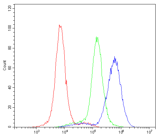
ARG42583 anti-RAB27A antibody FACS image
Flow Cytometry: K562 cells were blocked with 10% normal goat serum and then stained with ARG42583 anti-RAB27A antibody (blue) at 1 µg/10^6 cells for 30 min at 20°C, followed by incubation with DyLight®488 labelled secondary antibody. Isotype control antibody (green) was Rabbit IgG (1 µg/10^6 cells) used under the same conditions. Unlabelled sample (red) was also used as a control.
-

ARG42583 anti-RAB27A antibody IHC-P image
Immunohistochemistry: Paraffin-embedded Human mammary cancer tissue. Antigen Retrieval: Heat mediation was performed in Citrate buffer (pH 6.0) for 20 min. The tissue section was blocked with 10% goat serum. The tissue section was then stained with ARG42583 anti-RAB27A antibody at 1 µg/ml dilution, overnight at 4°C.
-

ARG42583 anti-RAB27A antibody IHC-P image
Immunohistochemistry: Paraffin-embedded Rat spleen tissue. Antigen Retrieval: Heat mediation was performed in Citrate buffer (pH 6.0) for 20 min. The tissue section was blocked with 10% goat serum. The tissue section was then stained with ARG42583 anti-RAB27A antibody at 1 µg/ml dilution, overnight at 4°C.
-

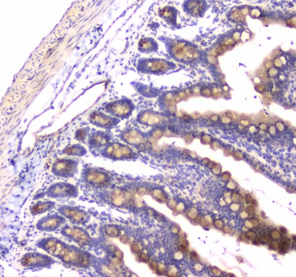
ARG42583 anti-RAB27A antibody IHC-P image
Immunohistochemistry: Paraffin-embedded Rat small intestine tissue. Antigen Retrieval: Heat mediation was performed in Citrate buffer (pH 6.0) for 20 min. The tissue section was blocked with 10% goat serum. The tissue section was then stained with ARG42583 anti-RAB27A antibody at 1 µg/ml dilution, overnight at 4°C.
-

ARG42583 anti-RAB27A antibody IHC-P image
Immunohistochemistry: Paraffin-embedded Mouse spleen tissue. Antigen Retrieval: Heat mediation was performed in Citrate buffer (pH 6.0) for 20 min. The tissue section was blocked with 10% goat serum. The tissue section was then stained with ARG42583 anti-RAB27A antibody at 1 µg/ml dilution, overnight at 4°C.