ARG42833
anti-R Cadherin / Cadherin 4 antibody
anti-R Cadherin / Cadherin 4 antibody for Flow cytometry,IHC-Formalin-fixed paraffin-embedded sections,Western blot and Human
Overview
| Product Description | Rabbit Polyclonal antibody recognizes R Cadherin / Cadherin 4 |
|---|---|
| Tested Reactivity | Hu |
| Tested Application | FACS, IHC-P, WB |
| Host | Rabbit |
| Clonality | Polyclonal |
| Isotype | IgG |
| Target Name | R Cadherin / Cadherin 4 |
| Antigen Species | Human |
| Immunogen | Recombinant protein corresponding to K458-D641 of Human R Cadherin / Cadherin 4. |
| Conjugation | Un-conjugated |
| Alternate Names | R-CAD; Cadherin-4; Retinal cadherin; CAD4; RCAD; R-cadherin |
Application Instructions
| Application Suggestion |
|
||||||||
|---|---|---|---|---|---|---|---|---|---|
| Application Note | IHC-P: Antigen Retrieval: Heat mediation was performed in EDTA buffer (pH 8.0). * The dilutions indicate recommended starting dilutions and the optimal dilutions or concentrations should be determined by the scientist. |
||||||||
| Observed Size | 130 kDa |
Properties
| Form | Liquid |
|---|---|
| Purification | Affinity purification with immunogen. |
| Buffer | 0.2% Na2HPO4, 0.9% NaCl, 0.05% Sodium azide and 4% Trehalose. |
| Preservative | 0.05% Sodium azide |
| Stabilizer | 4% Trehalose |
| Concentration | 0.5 mg/ml |
| Storage Instruction | For continuous use, store undiluted antibody at 2-8°C for up to a week. For long-term storage, aliquot and store at -20°C or below. Storage in frost free freezers is not recommended. Avoid repeated freeze/thaw cycles. Suggest spin the vial prior to opening. The antibody solution should be gently mixed before use. |
| Note | For laboratory research only, not for drug, diagnostic or other use. |
Bioinformation
| Database Links | |
|---|---|
| Gene Symbol | CDH4 |
| Gene Full Name | cadherin 4, type 1, R-cadherin (retinal) |
| Background | This gene is a classical cadherin from the cadherin superfamily. The encoded protein is a calcium-dependent cell-cell adhesion glycoprotein comprised of five extracellular cadherin repeats, a transmembrane region and a highly conserved cytoplasmic tail. Based on studies in chicken and mouse, this cadherin is thought to play an important role during brain segmentation and neuronal outgrowth. In addition, a role in kidney and muscle development is indicated. Of particular interest are studies showing stable cis-heterodimers of cadherins 2 and 4 in cotransfected cell lines. Previously thought to interact in an exclusively homophilic manner, this is the first evidence of cadherin heterodimerization. Three transcript variants encoding different isoforms have been found for this gene. [provided by RefSeq, Nov 2011] |
| Function | Cadherins are calcium-dependent cell adhesion proteins. They preferentially interact with themselves in a homophilic manner in connecting cells; cadherins may thus contribute to the sorting of heterogeneous cell types. May play an important role in retinal development. [UniProt] |
| Cellular Localization | Cell membrane; Single-pass type I membrane protein. [UniProt] |
| Calculated MW | 100 kDa |
Images (3) Click the Picture to Zoom In
-
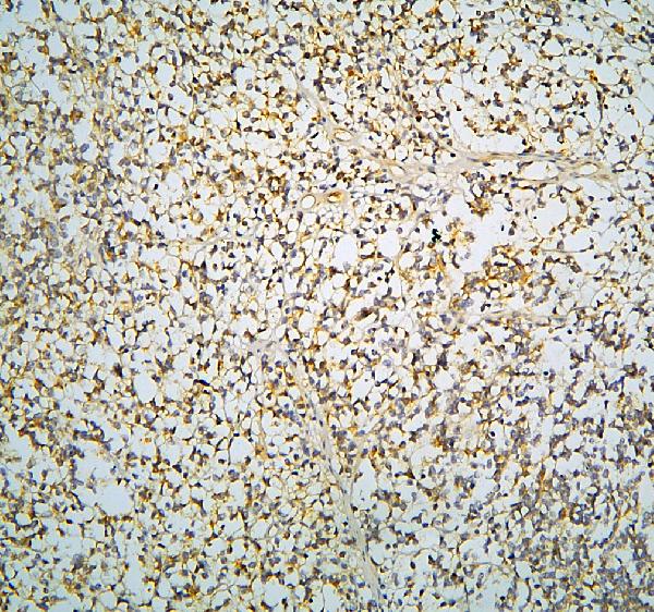
ARG42833 anti-R Cadherin / Cadherin 4 antibody IHC-P image
Immunohistochemistry: Paraffin-embedded Human glioma tissue. Antigen Retrieval: Heat mediation was performed in EDTA buffer (pH 8.0). The tissue section was blocked with 10% goat serum. The tissue section was then stained with ARG42833 anti-R Cadherin / Cadherin 4 antibody at 1 µg/ml dilution, overnight at 4°C.
-

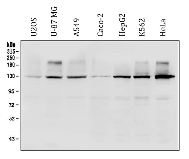
ARG42833 anti-R Cadherin / Cadherin 4 antibody WB image
Western blot: 50 µg of sample under reducing conditions. U2OS, U-87 MG, A549, Caco-2, HepG2, K562 and HeLa whole cell lysates stained with ARG42833 anti-R Cadherin / Cadherin 4 antibody at 0.5 µg/ml dilution, overnight at 4°C.
-
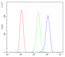
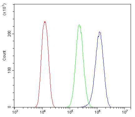
ARG42833 anti-R Cadherin / Cadherin 4 antibody FACS image
Flow Cytometry: U2OS cells were blocked with 10% normal goat serum and then stained with ARG42833 anti-R Cadherin / Cadherin 4 antibody (blue) at 1 µg/10^6 cells for 30 min at 20°C, followed by incubation with DyLight®488 labelled secondary antibody. Isotype control antibody (green) was Rabbit IgG (1 µg/10^6 cells) used under the same conditions. Unlabelled sample (red) was also used as a control.