ARG55619
anti-Pyk 2 antibody
anti-Pyk 2 antibody for Western blot and Mouse,Rat
Cancer antibody; Metabolism antibody; Signaling Transduction antibody
Overview
| Product Description | Rabbit Polyclonal antibody recognizes Pyk 2 |
|---|---|
| Tested Reactivity | Ms, Rat |
| Tested Application | WB |
| Host | Rabbit |
| Clonality | Polyclonal |
| Isotype | IgG |
| Target Name | Pyk 2 |
| Antigen Species | Mouse |
| Immunogen | KLH-conjugated synthetic peptide corresponding to aa. 769-802 of Mouse Pyk 2. |
| Conjugation | Un-conjugated |
| Alternate Names | FAK2; Proline-rich tyrosine kinase 2; Focal adhesion kinase 2; FADK2; RAFTK; Calcium-dependent tyrosine kinase; PYK2; FADK 2; CAK-beta; CADTK; Cell adhesion kinase beta; E430023O05Rik; Protein-tyrosine kinase 2-beta; Calcium-regulated non-receptor proline-rich tyrosine kinase; Raftk; EC 2.7.10.2; Related adhesion focal tyrosine kinase; CAKbeta; CAKB |
Application Instructions
| Application Suggestion |
|
||||
|---|---|---|---|---|---|
| Application Note | * The dilutions indicate recommended starting dilutions and the optimal dilutions or concentrations should be determined by the scientist. | ||||
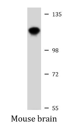
| Positive Control | Mouse brain |
Properties
| Form | Liquid |
|---|---|
| Purification | Purification with Protein A and immunogen peptide. |
| Buffer | PBS and 0.09% (W/V) Sodium azide |
| Preservative | 0.09% (W/V) Sodium azide |
| Storage Instruction | For continuous use, store undiluted antibody at 2-8°C for up to a week. For long-term storage, aliquot and store at -20°C or below. Storage in frost free freezers is not recommended. Avoid repeated freeze/thaw cycles. Suggest spin the vial prior to opening. The antibody solution should be gently mixed before use. |
| Note | For laboratory research only, not for drug, diagnostic or other use. |
Bioinformation
| Database Links | |
|---|---|
| Gene Symbol | Ptk2b |
| Gene Full Name | PTK2 protein tyrosine kinase 2 beta |
| Function | Non-receptor protein-tyrosine kinase that regulates reorganization of the actin cytoskeleton, cell polarization, cell migration, adhesion, spreading and bone remodeling. Plays a role in the regulation of the humoral immune response, and is required for normal levels of marginal B-cells in the spleen and normal migration of splenic B-cells. Required for normal macrophage polarization and migration towards sites of inflammation. Regulates cytoskeleton rearrangement and cell spreading in T-cells, and contributes to the regulation of T-cell responses. Promotes osteoclastic bone resorption; this requires both PTK2B/PYK2 and SRC. May inhibit differentiation and activity of osteoprogenitor cells. Functions in signaling downstream of integrin and collagen receptors, immune receptors, G-protein coupled receptors (GPCR), cytokine, chemokine and growth factor receptors, and mediates responses to cellular stress. Forms multisubunit signaling complexes with SRC and SRC family members upon activation; this leads to the phosphorylation of additional tyrosine residues, creating binding sites for scaffold proteins, effectors and substrates. Regulates numerous signaling pathways. Promotes activation of phosphatidylinositol 3-kinase and of the AKT1 signaling cascade. Promotes activation of NOS3. Regulates production of the cellular messenger cGMP. Promotes activation of the MAP kinase signaling cascade, including activation of MAPK1/ERK2, MAPK3/ERK1 and MAPK8/JNK1. Promotes activation of Rho family GTPases, such as RHOA and RAC1. Recruits the ubiquitin ligase MDM2 to P53/TP53 in the nucleus, and thereby regulates P53/TP53 activity, P53/TP53 ubiquitination and proteasomal degradation. Acts as a scaffold, binding to both PDPK1 and SRC, thereby allowing SRC to phosphorylate PDPK1 at 'Tyr-9, 'Tyr-373', and 'Tyr-376' (By similarity). Promotes phosphorylation of NMDA receptors by SRC family members, and thereby contributes to the regulation of NMDA receptor ion channel activity and intracellular Ca(2+) levels. May also regulate potassium ion transport by phosphorylation of potassium channel subunits. Phosphorylates SRC; this increases SRC kinase activity. Phosphorylates ASAP1, NPHP1, KCNA2 and SHC1. Promotes phosphorylation of ASAP2, RHOU and PXN; this requires both SRC and PTK2/PYK2 (By similarity). [UniProt] |
| Cellular Localization | Cytoplasm. Cytoplasm, perinuclear region. Cell membrane; Peripheral membrane protein; Cytoplasmic side. Cell junction, focal adhesion. Cell projection, lamellipodium. Cytoplasm, cell cortex Nucleus. Note=Colocalizes with integrins at the cell periphery (By similarity). Interaction with NPHP1 induces the membrane-association of the kinase. Colocalizes with PXN at the microtubule-organizing center. The tyrosine phosphorylated form is detected at cell-cell contacts. |
| Research Area | Cancer antibody; Metabolism antibody; Signaling Transduction antibody |
| Calculated MW | 116 kDa |
| PTM | Phosphorylated on tyrosine residues in response to various stimuli that elevate the intracellular calcium concentration; this activation is indirect and may be mediated by production of reactive oxygen species (ROS). Tyr-402 is the major autophosphorylation site, but other kinases can also phosphorylate Tyr-402. Autophosphorylation occurs in trans, i.e. one subunit of the dimeric receptor phosphorylates tyrosine residues on the other subunit. Phosphorylation at Tyr-402 promotes interaction with SRC and SRC family members, leading to phosphorylation at Tyr-579; Tyr-580 and Tyr-881. Phosphorylation at Tyr-881 is important for interaction with GRB2. Phosphorylated on tyrosine residues upon activation of FGR and PKC. Recruitment by NPHP1 to cell matrix adhesions initiates Tyr-402 phosphorylation. In monocytes, adherence to substrata is required for tyrosine phosphorylation and kinase activation. Angiotensin II, thapsigargin and L-alpha-lysophosphatidic acid (LPA) also induce autophosphorylation and increase kinase activity. Phosphorylation by MYLK promotes ITGB2 activation and is thus essential to trigger neutrophil transmigration during lung injury. Dephosphorylated by PTPN12. |
Images (1) Click the Picture to Zoom In
-
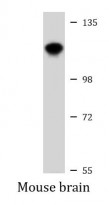
ARG55619 anti-Pyk 2 antibody WB image
Western blot: 20 µg of Mouse brain lysate stained with ARG55619 anti-Pyk 2 antibody at 1:1000 dilution.