ARG43992
anti-Proteasome 20S LMP2 antibody
anti-Proteasome 20S LMP2 antibody for ELISA,Flow cytometry,IHC-Formalin-fixed paraffin-embedded sections,Western blot and Human,Mouse,Rat
Overview
| Product Description | Rabbit Polyclonal antibody recognizes Proteasome 20S LMP2 |
|---|---|
| Tested Reactivity | Hu, Ms, Rat |
| Tested Application | ELISA, FACS, IHC-P, WB |
| Host | Rabbit |
| Clonality | Polyclonal |
| Isotype | IgG |
| Target Name | Proteasome 20S LMP2 |
| Antigen Species | Human |
| Immunogen | Human Proteasome 20S LMP2 recombinant protein |
| Expression System | E.coli |
| Conjugation | Un-conjugated |
| Protein Full Name | Proteasome subunit beta type-9 |
| Alternate Names | PSMB9; Proteasome 20S Subunit Beta 9; RING12; PSMB6i; Beta1i; LMP2 ; Multicatalytic Endopeptidase Complex Chain 7; Really Interesting New Gene 12 Protein; Large Multifunctional Peptidase 2; Proteasome Subunit Beta Type-9; Low Molecular Mass Protein 2; Proteasome Subunit Beta-1i; Proteasome Subunit Beta 9; Proteasome Chain 7; Macropain Chain 7; EC 3.4.25.1; Proteasome (Prosome, Macropain) Subunit, Beta Type, 9 ; Proteasome Catalytic Subunit 1i; Proteasome Subunit Beta 6i; Proteasome Subunit Beta1i; Proteasome-Related Gene; Proteasome Subunit Β1i; PRAAS3 |
Application Instructions
| Application Suggestion |
|
||||||||||
|---|---|---|---|---|---|---|---|---|---|---|---|
| Application Note | * The dilutions indicate recommended starting dilutions and the optimal dilutions or concentrations should be determined by the scientist. |
Properties
| Form | Liquid |
|---|---|
| Purification | Affinity purified with Immunogen. |
| Buffer | 0.9% NaCl, 0.2% Na2HPO4 and 4% Trehalose. |
| Stabilizer | 4% Trehalose |
| Concentration | 0.5 mg/ml |
| Storage Instruction | For continuous use, store undiluted antibody at 2-8°C for up to a week. For long-term storage, aliquot and store at -20°C or below. Storage in frost free freezers is not recommended. Avoid repeated freeze/thaw cycles. Suggest spin the vial prior to opening. The antibody solution should be gently mixed before use. |
| Note | For laboratory research only, not for drug, diagnostic or other use. |
Bioinformation
| Database Links | |
|---|---|
| Gene Symbol | PSMB9 |
| Gene Full Name | Proteasome 20S Subunit Beta 9 |
| Background | The proteasome is a multicatalytic proteinase complex with a highly ordered ring-shaped 20S core structure. The core structure is composed of 4 rings of 28 non-identical subunits; 2 rings are composed of 7 alpha subunits and 2 rings are composed of 7 beta subunits. Proteasomes are distributed throughout eukaryotic cells at a high concentration and cleave peptides in an ATP/ubiquitin-dependent process in a non-lysosomal pathway. An essential function of a modified proteasome, the immunoproteasome, is the processing of class I MHC peptides. This gene encodes a member of the proteasome B-type family, also known as the T1B family, that is a 20S core beta subunit. This gene is located in the class II region of the MHC (major histocompatibility complex). Expression of this gene is induced by gamma interferon and this gene product replaces catalytic subunit 1 (proteasome beta 6 subunit) in the immunoproteasome. Proteolytic processing is required to generate a mature subunit. |
| Function | The proteasome is a multicatalytic proteinase complex which is characterized by its ability to cleave peptides with Arg, Phe, Tyr, Leu, and Glu adjacent to the leaving group at neutral or slightly basic pH. The proteasome has an ATP-dependent proteolytic activity. This subunit is involved in antigen processing to generate class I binding peptides. Replacement of PSMB6 by PSMB9 increases the capacity of the immunoproteasome to cleave model peptides after hydrophobic and basic residues. |
| Cellular Localization | Cytoplasm, Nucleus, Proteasome |
| Calculated MW | 23 kDa |
| PTM | Acetylation, Zymogen |
Images (5) Click the Picture to Zoom In
-
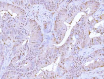
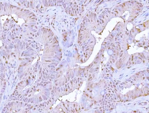
ARG43992 anti-Proteasome 20S LMP2 antibody IHC-P image
Immunohistochemistry: Humanrectum adenocarcinoma stained with ARG43992 anti-Proteasome 20S LMP2 antibody at 2 μg/ml dilution.
-
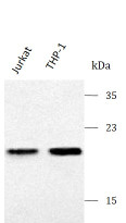
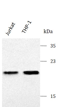
ARG43992 anti-Proteasome 20S LMP2 antibody WB image
Western blot:Jurkat and THP-1 stained with ARG43992 anti-Proteasome 20S LMP2 antibody at 0.5 μg/mL dilution.
-
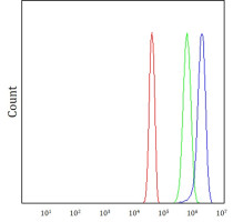
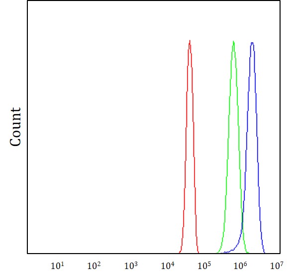
ARG43992 anti-Proteasome 20S LMP2 antibody FACS image
Flow Cytometry: JK cells stained withARG43992 anti-Proteasome 20S LMP2 antibody (blue) at 1 μg/1x10^6 cells dilution.
-
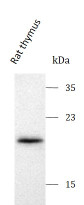
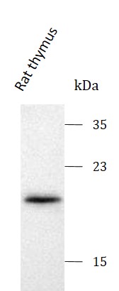
ARG43992 anti-Proteasome 20S LMP2 antibody WB image
Western blot: Rat thymus ARG43992 anti-Proteasome 20S LMP2 antibody at 0.5 μg/mL dilution.
-
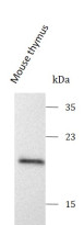
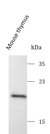
ARG43992 anti-Proteasome 20S LMP2 antibody WB image
Western blot: Mouse thymus ARG43992 anti-Proteasome 20S LMP2 antibody at 0.5 μg/mL dilution.