ARG58969
anti-Periplakin antibody
anti-Periplakin antibody for ICC/IF,Western blot and Human,Mouse,Rat
Overview
| Product Description | Rabbit Polyclonal antibody recognizes Periplakin |
|---|---|
| Tested Reactivity | Hu, Ms, Rat |
| Tested Application | ICC/IF, WB |
| Host | Rabbit |
| Clonality | Polyclonal |
| Isotype | IgG |
| Target Name | Periplakin |
| Antigen Species | Human |
| Immunogen | Recombinant fusion protein corresponding to aa. 1-130 of Human Periplakin (NP_002696.3). |
| Conjugation | Un-conjugated |
| Alternate Names | 190 kDa paraneoplastic pemphigus antigen; Periplakin; 195 kDa cornified envelope precursor protein |
Application Instructions
| Application Suggestion |
|
||||||
|---|---|---|---|---|---|---|---|
| Application Note | * The dilutions indicate recommended starting dilutions and the optimal dilutions or concentrations should be determined by the scientist. | ||||||
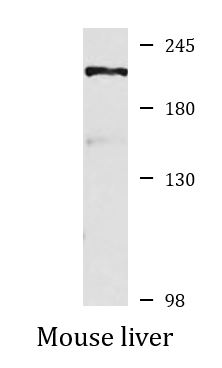
| Positive Control | Mouse liver | ||||||
| Observed Size | 205 kDa |
Properties
| Form | Liquid |
|---|---|
| Purification | Affinity purified. |
| Buffer | PBS (pH 7.3), 0.02% Sodium azide and 50% Glycerol. |
| Preservative | 0.02% Sodium azide |
| Stabilizer | 50% Glycerol |
| Storage Instruction | For continuous use, store undiluted antibody at 2-8°C for up to a week. For long-term storage, aliquot and store at -20°C. Storage in frost free freezers is not recommended. Avoid repeated freeze/thaw cycles. Suggest spin the vial prior to opening. The antibody solution should be gently mixed before use. |
| Note | For laboratory research only, not for drug, diagnostic or other use. |
Bioinformation
| Database Links | |
|---|---|
| Gene Symbol | PPL |
| Gene Full Name | periplakin |
| Background | The protein encoded by this gene is a component of desmosomes and of the epidermal cornified envelope in keratinocytes. The N-terminal domain of this protein interacts with the plasma membrane and its C-terminus interacts with intermediate filaments. Through its rod domain, this protein forms complexes with envoplakin. This protein may serve as a link between the cornified envelope and desmosomes as well as intermediate filaments. AKT1/PKB, a protein kinase mediating a variety of cell growth and survival signaling processes, is reported to interact with this protein, suggesting a possible role for this protein as a localization signal in AKT1-mediated signaling. [provided by RefSeq, Jul 2008] |
| Function | Component of the cornified envelope of keratinocytes. May link the cornified envelope to desmosomes and intermediate filaments. May act as a localization signal in PKB/AKT-mediated signaling. [UniProt] |
| Cellular Localization | Cell junction, desmosome, Cytoplasm, cytoskeleton, Cell membrane, Nucleus, Mitochondrion. [UniProt] |
| Calculated MW | 205 kDa |
Images (2) Click the Picture to Zoom In
-

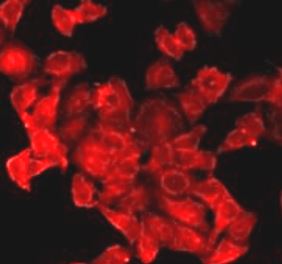
ARG58969 anti-Periplakin antibody ICC/IF image
Immunofluorescence: MCF7 cells stained with ARG58969 anti-Periplakin antibody.
-
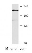
ARG58969 anti-Periplakin antibody WB image
Western blot: 25 µg of Mouse liver lysate stained with ARG58969 anti-Periplakin antibody at 1:1000 dilution.