ARG52395
anti-Peripherin antibody
anti-Peripherin antibody for ICC/IF,Western blot and Mouse,Rat
Neuroscience antibody; Signaling Transduction antibody
Overview
| Product Description | Chicken Polyclonal antibody recognizes Peripherin |
|---|---|
| Tested Reactivity | Ms, Rat |
| Predict Reactivity | Hu, Bov, Pig |
| Tested Application | ICC/IF, WB |
| Host | Chicken |
| Clonality | Polyclonal |
| Isotype | IgY |
| Target Name | Peripherin |
| Antigen Species | Rat |
| Immunogen | Recombinant rat peripherin expressed in and purified from E. coli |
| Conjugation | Un-conjugated |
| Alternate Names | Peripherin; NEF4; PRPH1; Neurofilament 4 |
Application Instructions
| Application Suggestion |
|
||||||
|---|---|---|---|---|---|---|---|
| Application Note | Specific for the ~57kDa peripherin protein. * The dilutions indicate recommended starting dilutions and the optimal dilutions or concentrations should be determined by the scientist. |
||||||
| Positive Control | Rat cerebellar lysate | ||||||
| Observed Size | ~ 57 kDa |
Properties
| Form | Liquid |
|---|---|
| Purification | Total IgY fraction |
| Buffer | Total IgY fraction in PBS and 10 mM Sodium azide |
| Preservative | 10 mM Sodium azide |
| Storage Instruction | For continuous use, store undiluted antibody at 2-8°C for up to a week. For long-term storage, aliquot and store at -20°C or below. Storage in frost free freezers is not recommended. Avoid repeated freeze/thaw cycles. Suggest spin the vial prior to opening. The antibody solution should be gently mixed before use. |
| Note | For laboratory research only, not for drug, diagnostic or other use. |
Bioinformation
| Database Links | |
|---|---|
| Background | Peripherin is a ~57kDa intermediate filament subunit found initially in sensory neurons of the peripheral nervous systems, which gives the protein its name. Subsequently, peripherin was found in some sensory and other neurons of the central nervous system and also in PC12 cells. Peripherin is also expressed in certain neuroendocrine tumors and in the insulin producing cells of the pancreas. Peripherin belongs to the Class III family of intermediate filament subunits which also includes vimentin, glial fibrillary acidic protein (GFAP) and desmin. In contrast to the neurofilaments, peripherin is strongly up-regulated after nerve injury . Antibodies to peripherin can be used in identifying, classifying, and studying neurons throughout the nervous system. Peripherin is also a good diagnostic marker for ballooned axons seen in Lou Gehrig's disease (Amyotrophic lateral sclerosis) and some neuronally derived tumors . Autoantibodies to peripherin are frequently seen in the sera of patients with diabetes . Peripherin is not related to peripherin/RDS, a protein of the photoreceptor outer membrane mutations of which are causative of certain forms of slow retinal degeneration. |
| Research Area | Neuroscience antibody; Signaling Transduction antibody |
| Calculated MW | 54 kDa |
Images (4) Click the Picture to Zoom In
-
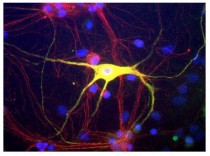
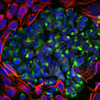
ARG52395 anti-Peripherin antibody ICC/IF image
Immunofluorescence: Cultured Rat neurons and glia stained with ARG52395 anti-Peripherin antibody showing peripherin in green and alpha-internexin in red. Cells expressing both peripherin and alpha-internexin appear yellow.
-
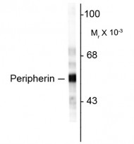
ARG52395 anti-Peripherin antibody WB image
Western blot: Rat cerebellar lysate stained with ARG52395 anti-Peripherin antibody.
-
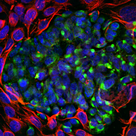
ARG52395 anti-Peripherin antibody ICC/IF image
Immunofluorescence: Mixed fibroblast and PC-12 pheochromocytoma cell culture stained with ARG52395 anti-Peripherin antibody (green) at 1:5000 dilution, and costained with anti-Vimentin antibody (red) at 1:2000 dilution. DAPI (blue) for nuclear staining.
The Peripherin antibody reveals cytoplasmic filamentous staining in the small PC-12 cells, while Vimentin antibody stains intermediate filaments in the surrounding fibroblastic cells which do not express Peripherin.
-
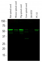
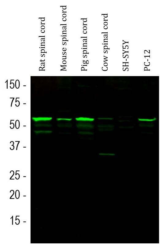
ARG52395 anti-Peripherin antibody WB image
Western blot: Rat spinal cord, Mouse spinal cord, Pig spinal cord, Cow spinal cord, SH-SY5Y and PC-12 cell lysates stained with ARG52395 anti-Peripherin antibody (green) at 1:10000 dilution.