ARG10726
anti-Peripherin antibody
anti-Peripherin antibody for ICC/IF,IHC-Frozen sections,Western blot and Human,Mouse,Rat,Cow,Pig
Overview
| Product Description | Rabbit Polyclonal antibody recognizes Peripherin |
|---|---|
| Tested Reactivity | Hu, Ms, Rat, Cow, Pig |
| Tested Application | ICC/IF, IHC-Fr, WB |
| Host | Rabbit |
| Clonality | Polyclonal |
| Isotype | IgG |
| Target Name | Peripherin |
| Antigen Species | Human |
| Immunogen | Recombinant Human peripherin expressed in and purified from E. coli. |
| Conjugation | Un-conjugated |
| Alternate Names | Peripherin; NEF4; PRPH1; Neurofilament 4 |
Application Instructions
| Application Suggestion |
|
||||||||
|---|---|---|---|---|---|---|---|---|---|
| Application Note | * The dilutions indicate recommended starting dilutions and the optimal dilutions or concentrations should be determined by the scientist. |
Properties
| Form | Liquid |
|---|---|
| Purification | Unpurified. |
| Buffer | Serum. |
| Storage Instruction | For continuous use, store undiluted antibody at 2-8°C for up to a week. For long-term storage, aliquot and store at -20°C or below. Storage in frost free freezers is not recommended. Avoid repeated freeze/thaw cycles. Suggest spin the vial prior to opening. The antibody solution should be gently mixed before use. |
| Note | For laboratory research only, not for drug, diagnostic or other use. |
Bioinformation
| Database Links | |
|---|---|
| Gene Symbol | PRPH |
| Gene Full Name | peripherin |
| Background | This gene encodes a cytoskeletal protein found in neurons of the peripheral nervous system. The encoded protein is a type III intermediate filament protein with homology to other cytoskeletal proteins such as desmin, and is a different protein that the peripherin found in photoreceptors. Mutations in this gene have been associated with susceptibility to amyotrophic lateral sclerosis. [provided by RefSeq, Jul 2008] |
| Function | Class-III neuronal intermediate filament protein. [UniProt] |
| Calculated MW | 54 kDa |
Images (4) Click the Picture to Zoom In
-

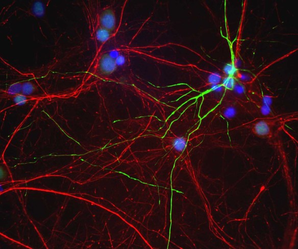
ARG10726 anti-Peripherin antibody ICC/IF image
Immunocytochemistry: Mixed neuron / glia cultures from newborn Rat brain stained with ARG10726 anti-Peripherin antibody (green) and co-stained with chicken polyclonal antibody to NF-H (red). A class of large neurons, like the one at the top right of this image, contain peripherin, while the majority of neurons and their processes contain NF-H and not peripherin. The blue channel reveals the localization of DNA.
-
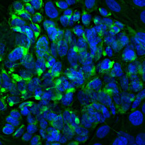
ARG10726 anti-Peripherin antibody ICC/IF image
Immunofluorescence: PC-12 cells stained with ARG10726 anti-Peripherin antibody (green) at 1:2000 dilution. Hoechst (blue) for nuclear staining.
Peripherin, one of the Class III family of intermediate filament subunit proteins, is a major component of the PC-12 cell forming a perinuclear cap, with some filaments visible in the cytoplasm.
-
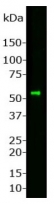
ARG10726 anti-Peripherin antibody WB image
Western blot: Whole Rat spinal cord homogenate stained with ARG10726 anti-Peripherin antibody at 1:1000 dilution. A prominent band running with an apparent SDS-PAGE molecular weight of ~57 kDa corresponds to Peripherin.
-
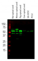
ARG10726 anti-Peripherin antibody WB image
Western blot: Rat spinal cord, Mouse spinal cord, Pig spinal cord, Cow spinal cord, SH-SY5Y and PC12 cell lysates stained with ARG10726 anti-Peripherin antibody (green) at 1:10000 dilution.
The major band at ~ 57 kDa corresponds to the major peripherin protein isoform, while other bands presumably represent protein products of alternate transcripts of the peripherin gene.