ARG52394
anti-Peripherin antibody [7C5]
anti-Peripherin antibody [7C5] for ICC/IF,Western blot and Mouse,Rat
Neuroscience antibody; Signaling Transduction antibody
Overview
| Product Description | Mouse Monoclonal antibody [7C5] recognizes Peripherin |
|---|---|
| Tested Reactivity | Ms, Rat |
| Tested Application | ICC/IF, WB |
| Host | Mouse |
| Clonality | Monoclonal |
| Clone | 7C5 |
| Isotype | IgG1 |
| Target Name | Peripherin |
| Antigen Species | Rat |
| Immunogen | Recombinant rat peripherin expressed in and purified from E. coli |
| Conjugation | Un-conjugated |
| Alternate Names | Peripherin; NEF4; PRPH1; Neurofilament 4 |
Application Instructions
| Application Suggestion |
|
||||||
|---|---|---|---|---|---|---|---|
| Application Note | Specific for the ~57kDa peripherin protein. This antibody performs well on aldehyde fixed tissues. * The dilutions indicate recommended starting dilutions and the optimal dilutions or concentrations should be determined by the scientist. |
Properties
| Form | Liquid |
|---|---|
| Purification | Total IgG fraction |
| Buffer | Total IgG fraction and 10 mM Sodium azide |
| Preservative | 10 mM Sodium azide |
| Storage Instruction | For continuous use, store undiluted antibody at 2-8°C for up to a week. For long-term storage, aliquot and store at -20°C or below. Storage in frost free freezers is not recommended. Avoid repeated freeze/thaw cycles. Suggest spin the vial prior to opening. The antibody solution should be gently mixed before use. |
| Note | For laboratory research only, not for drug, diagnostic or other use. |
Bioinformation
| Database Links | |
|---|---|
| Background | Peripherin is a ~57kDa intermediate filament subunit found initially in sensory neurons of the peripheral nervous systems, which gives the protein its name. Subsequently, peripherin was found in some sensory and other neurons of the central nervous system and also in PC12 cells. Peripherin is also expressed in certain neuroendocrine tumors and in the insulin producing cells of the pancreas. Peripherin belongs to the Class III family of intermediate filament subunits which also includes vimentin, glial fibrillary acidic protein (GFAP) and desmin. In contrast to the neurofilaments, peripherin is strongly up-regulated after nerve injury . Antibodies to peripherin can be used in identifying, classifying, and studying neurons throughout the nervous system. Peripherin is also a good diagnostic marker for ballooned axons seen in Lou Gehrig's disease (Amyotrophic lateral sclerosis) and some neuronally derived tumors . Autoantibodies to peripherin are frequently seen in the sera of patients with diabetes . Peripherin is not related to peripherin/RDS, a protein of the photoreceptor outer membrane mutations of which are causative of certain forms of slow retinal degeneration. |
| Research Area | Neuroscience antibody; Signaling Transduction antibody |
| Calculated MW | 54 kDa |
Images (4) Click the Picture to Zoom In
-
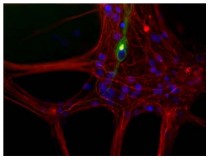
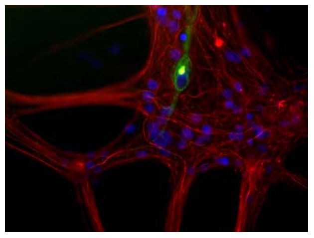
ARG52394 anti-Peripherin antibody [7C5] ICC/IF image
Immunofluorescence: Cultured newborn rat neurons and glia stained with ARG52394 anti-Peripherin antibody [7C5] showing peripherin in green and neurofilament L in red.
-
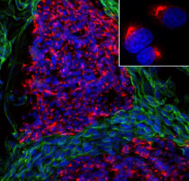
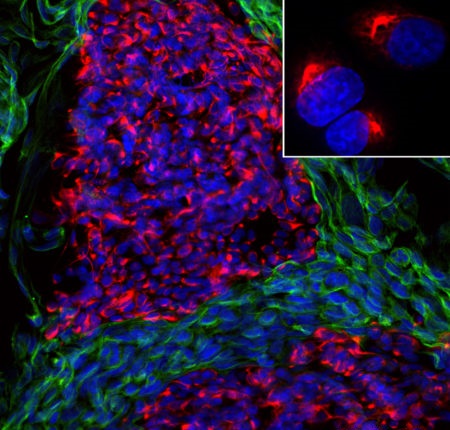
ARG52394 anti-Peripherin antibody [7C5] ICC/IF image
Immunofluorescence: Mixed fibroblast and Rat pheochromocytoma PC-12 cell culture stained with ARG52394 anti-Peripherin antibody [7C5] (red) at 1:500 dilution, and costained with ARG52468 anti-Vimentin antibody (green) at 1:10000 dilution. Hoechst (blue) for nuclear staining.
Clone 7C5 reveals cytoplasmic filamentous staining in the small PC-12 cells, while Vimentin antibody stains intermediate filaments in the surrounding fibroblastic cells which do not express Peripherin.
-
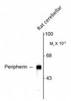
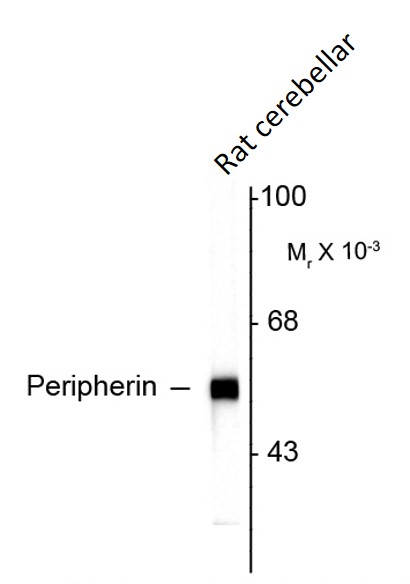
ARG52394 anti-Peripherin antibody [7C5] WB image
Western blot: Rat cerebellar lysate showing specific immunolabeling of the ~ 57 kDa peripherin protein stained with ARG52394 anti-Peripherin antibody [7C5].
-
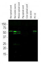
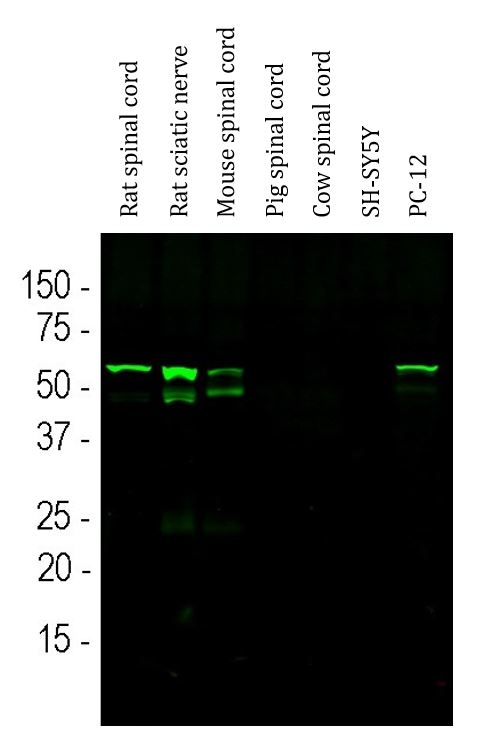
ARG52394 anti-Peripherin antibody [7C5] WB image
Western blot: Rat spinal cord, Rat sciatic nerve, Mouse spinal cord, Pig spinal cord, Cow spinal cord, SH-SY5Y and PC-12 cell lysates stained with ARG52394 anti-Peripherin antibody [7C5] (green) at 1:1000 dilution.
The band at ~ 57 kDa corresponds to the Peripherin protein detected only in Rat and Mouse lysates, since the antibody is rodent specific.
Clone References
Characterization of neurons from adult human skin-derived precursors in serum-free medium : a PCR array and immunocytological analysis.


Type III Nrg1 back signaling enhances functional TRPV1 along sensory axons contributing to basal and inflammatory thermal pain sensation.
IHC-P / Mouse
Peripheral nervous system progenitors can be reprogrammed to produce myelinating oligodendrocytes and repair brain lesions.
Phosphorylation of sodium channel Na(v)1.8 by p38 mitogen-activated protein kinase increases current density in dorsal root ganglion neurons.
Differential modulation of sodium channel Na(v)1.6 by two members of the fibroblast growth factor homologous factor 2 subfamily.


