ARG43855
anti-Perforin antibody [dG9] (PE-Cyanine 7)
anti-Perforin antibody [dG9] (PE-Cyanine 7) for Flow cytometry and Human
Overview
| Product Description | PE-Cyanine 7-conjugated Mouse Monoclonal antibody human Perforin |
|---|---|
| Tested Reactivity | Hu |
| Predict Reactivity | Cow |
| Tested Application | FACS |
| Host | Mouse |
| Clonality | Monoclonal |
| Clone | dG9 |
| Isotype | IgG2b kappa |
| Target Name | Perforin |
| Antigen Species | Human |
| Immunogen | Human YT lymphoma cell line |
| Conjugation | PE-Cyanine 7 |
| Protein Full Name | Perforin-1 |
| Alternate Names | PRF1; Perforin 1; PFP; P1; HPLH2; Perforin 1 (Pore Forming Protein); Lymphocyte Pore-Forming Protein; Perforin-1; Cytolysin; Perforin 1 (Preforming Protein); Perforin |
Application Instructions
| Application Suggestion |
|
||||
|---|---|---|---|---|---|
| Application Note | * The dilutions indicate recommended starting dilutions and the optimal dilutions or concentrations should be determined by the scientist. |
Properties
| Form | Liquid |
|---|---|
| Purification | Protein-A affinity chromatography |
| Buffer | PBS (pH 7.4) and 15 mM Sodium azide |
| Preservative | 15 mM Sodium azide |
| Storage Instruction | Aliquot and store in the dark at 4°C. Keep protected from prolonged exposure to light. Do not freeze. Suggest spin the vial prior to opening. The antibody solution should be gently mixed before use. |
Bioinformation
| Database Links | |
|---|---|
| Gene Symbol | PRF1 |
| Gene Full Name | Perforin 1 |
| Background | This gene encodes a protein with structural similarities to complement component C9 that is important in immunity. This protein forms membrane pores that allow the release of granzymes and subsequent cytolysis of target cells. Whether pore formation occurs in the plasma membrane of target cells or in an endosomal membrane inside target cells is subject to debate. Mutations in this gene are associated with a variety of human disease including diabetes, multiple sclerosis, lymphomas, autoimmune lymphoproliferative syndrome (ALPS), aplastic anemia, and familial hemophagocytic lymphohistiocytosis type 2 (FHL2), a rare and lethal autosomal recessive disorder of early childhood. |
| Function | Pore-forming protein that plays a key role in granzyme-mediated programmed cell death, and in defense against virus-infected or neoplastic cells. |
| Cellular Localization | Cell membrane, Endosome, Lysosome, Membrane, Secreted |
| Calculated MW | 61 kDa |
| PTM | Disulfide bond, Glycoprotein |
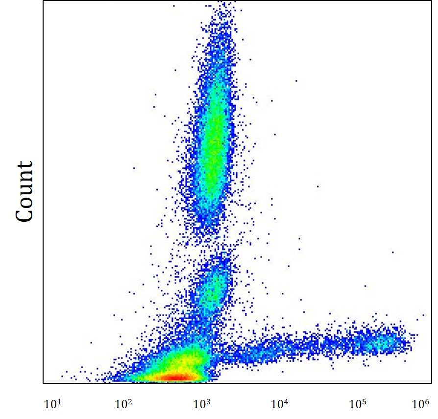
Images (1) Click the Picture to Zoom In
-

ARG43855 anti-Perforin antibody [dG9] (PE-Cyanine 7) FACS image
Flow Cytometry: Human whole blood stained with ARG43855 anti-Perforin antibody [dG9] (PE-Cyanine 7) at 4 μg.