ARG10725
anti-Parvalbumin antibody [3C9]
anti-Parvalbumin antibody [3C9] for ICC/IF,IHC-Frozen sections,Western blot and Human,Mouse,Rat,Cow
Overview
| Product Description | Mouse Monoclonal antibody [3C9] recognizes Parvalbumin |
|---|---|
| Tested Reactivity | Hu, Ms, Rat, Cow |
| Tested Application | ICC/IF, IHC-Fr, WB |
| Host | Mouse |
| Clonality | Monoclonal |
| Clone | 3C9 |
| Isotype | IgG1 |
| Target Name | Parvalbumin |
| Antigen Species | Human |
| Immunogen | Full-length recombinant Human Parvalbumin. |
| Conjugation | Un-conjugated |
| Alternate Names | PVALB; Parvalbumin; Parvalbumin Alpha; D22S749 |
Application Instructions
| Application Suggestion |
|
||||||||
|---|---|---|---|---|---|---|---|---|---|
| Application Note | * The dilutions indicate recommended starting dilutions and the optimal dilutions or concentrations should be determined by the scientist. |
Properties
| Form | Liquid |
|---|---|
| Purification | Affinity purification. |
| Concentration | 1 mg/ml |
| Storage Instruction | For continuous use, store undiluted antibody at 2-8°C for up to a week. For long-term storage, aliquot and store at -20°C or below. Storage in frost free freezers is not recommended. Avoid repeated freeze/thaw cycles. Suggest spin the vial prior to opening. The antibody solution should be gently mixed before use. |
| Note | For laboratory research only, not for drug, diagnostic or other use. |
Bioinformation
| Database Links | |
|---|---|
| Gene Symbol | PVALB |
| Gene Full Name | parvalbumin |
| Background | The protein encoded by this gene is a high affinity calcium ion-binding protein that is structurally and functionally similar to calmodulin and troponin C. The encoded protein is thought to be involved in muscle relaxation. Alternative splicing results in multiple transcript variants. |
| Function | In muscle, parvalbumin is thought to be involved in relaxation after contraction. It binds two calcium ions. |
| Calculated MW | 12 kDa |
| PTM | Acetylation, Phosphoprotein |
Images (4) Click the Picture to Zoom In
-
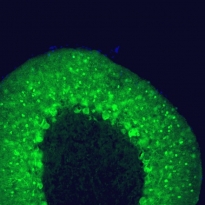
ARG10725 anti-Parvalbumin antibody [3C9] ICC/IF image
Immunocytochemistry: Adult Rat cerebellum floating section was stained with ARG10725 anti-Parvalbumin antibody [3C9] at 1:1000 (green). Parvalbumin is prominently expressed in the dendrites and perikarya of Purkinje cells and some interneurons in the molecular layer. Blue is a DNA stain.
-
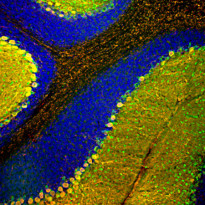
ARG10725 anti-Parvalbumin antibody [3C9] IHC-Fr image
Immunohistochemistry: Frozen section of Rat cerebellum stained with ARG10725 anti-Parvalbumin antibody [3C9] (green) at 1:1000 dilution and costained with Chicken pAb to calbindin (red) at 1:2000 dilution. DAPI (blue) for nuclear staining. (Sample preparation: Following transcardial perfusion of Rat with 4% paraformaldehyde, brain was post fixed for 24 hours, cut to 45 µM, and free-floating sections were stained with above antibodies.)
Most Purkinje cells coexpress both parvalbumin and calbindin and so appear yellow, whereas basket, stellate and Golgi cells express parvalbumin only and so appear green.
-
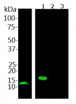
ARG10725 anti-Parvalbumin antibody [3C9] WB image
Western blot: Rat skeletal muscle lysate (left) and His-tagged recombinant proteins (right): 1) parvalbumin, 2) calretinin, and 3) calbindin. Blots were stained with ARG10725 anti-Parvalbumin antibody [3C9] at 1:1000. In skeletal muscle lysates, this antibody recognizes a band at 12 kDa which represents parvalbumin. It reacts to recombinant parvalbumin only, not the other calcium-binding proteins listed. Note, this antibody does not recognize parvabumin in Rat or mouse brain lysates on western blots. Protein from Rat skeletal muscle lysates were transferred to PVDF membrane.
-
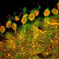
ARG10725 anti-Parvalbumin antibody [3C9] IHC-Fr image
Immunohistochemistry: Frozen section of Rat brain cerebellum stained with ARG10725 anti-Parvalbumin antibody [3C9] (green) at 1:1000 dilution and costained with Chicken pAb to calbindin (red) at 1:2000 dilution. (Sample preparation: Following transcardial perfusion of Rat with 4% paraformaldehyde, brain was post fixed for 24 hours, cut to 45 µM, and free-floating sections were stained with above antibodies.)
Most Purkinje cells coexpress both parvalbumin and calbindin and so appear yellow, whereas basket, stellate and Golgi cells express parvalbumin only and so appear green.


