ARG41973
anti-PTBP2 antibody
anti-PTBP2 antibody for Flow cytometry,ICC/IF,IHC-Formalin-fixed paraffin-embedded sections,Western blot and Human,Mouse,Rat
Overview
| Product Description | Rabbit Polyclonal antibody recognizes PTBP2 |
|---|---|
| Tested Reactivity | Hu, Ms, Rat |
| Tested Application | FACS, ICC/IF, IHC-P, WB |
| Host | Rabbit |
| Clonality | Polyclonal |
| Isotype | IgG |
| Target Name | PTBP2 |
| Antigen Species | Human |
| Immunogen | Recombinant protein corresponding to M1-A504 of Human PTBP2. |
| Conjugation | Un-conjugated |
| Alternate Names | Polypyrimidine tract-binding protein 2; nPTB; PTBLP; Neurally-enriched homolog of PTB; Neural polypyrimidine tract-binding protein; brPTB; PTB-like protein |
Application Instructions
| Application Suggestion |
|
||||||||||
|---|---|---|---|---|---|---|---|---|---|---|---|
| Application Note | IHC-P: Antigen Retrieval: Heat mediation was performed in Citrate buffer (pH 6.0, epitope retrieval solution) for 20 min. * The dilutions indicate recommended starting dilutions and the optimal dilutions or concentrations should be determined by the scientist. |
||||||||||
| Observed Size | 57 kDa |
Properties
| Form | Liquid |
|---|---|
| Purification | Affinity purification with immunogen. |
| Buffer | 0.2% Na2HPO4, 0.9% NaCl, 0.05% Sodium azide and 4% Trehalose. |
| Preservative | 0.05% Sodium azide |
| Stabilizer | 4% Trehalose |
| Concentration | 0.5 mg/ml |
| Storage Instruction | For continuous use, store undiluted antibody at 2-8°C for up to a week. For long-term storage, aliquot and store at -20°C or below. Storage in frost free freezers is not recommended. Avoid repeated freeze/thaw cycles. Suggest spin the vial prior to opening. The antibody solution should be gently mixed before use. |
| Note | For laboratory research only, not for drug, diagnostic or other use. |
Bioinformation
| Database Links | |
|---|---|
| Gene Symbol | PTBP2 |
| Gene Full Name | polypyrimidine tract binding protein 2 |
| Background | The protein encoded by this gene binds to intronic polypyrimidine clusters in pre-mRNA molecules and is implicated in controlling the assembly of other splicing-regulatory proteins. This protein is very similar to the polypyrimidine tract binding protein (PTB) but most of its isoforms are expressed primarily in the brain. Alternative splicing results in multiple transcript variants. [provided by RefSeq, Jul 2014] |
| Function | RNA-binding protein which binds to intronic polypyrimidine tracts and mediates negative regulation of exons splicing. May antagonize in a tissue-specific manner the ability of NOVA1 to activate exon selection. In addition to its function in pre-mRNA splicing, plays also a role in the regulation of translation. Isoform 5 has a reduced affinity for RNA. [UniProt] |
| Cellular Localization | Nucleus. [UniProt] |
| Calculated MW | 57 kDa |
Images (9) Click the Picture to Zoom In
-

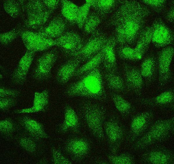
ARG41973 anti-PTBP2 antibody ICC/IF image
Immunofluorescence: U2OS cells were blocked with 10% goat serum and then stained with ARG41973 anti-PTBP2 antibody at 2 µg/ml dilution, overnight at 4°C.
-
ARG41973 anti-PTBP2 antibody IHC-P image
Immunohistochemistry: Paraffin-embedded Mouse cardiac muscle tissue. Antigen Retrieval: Heat mediation was performed in Citrate buffer (pH 6.0, epitope retrieval solution) for 20 min. The tissue section was blocked with 10% goat serum. The tissue section was then stained with ARG41973 anti-PTBP2 antibody at 1 µg/ml dilution, overnight at 4°C.
-
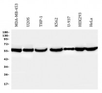
ARG41973 anti-PTBP2 antibody WB image
Western blot: 50 µg of samples under reducing conditions. MDA-MB-453, U2OS, THP-1, K562, U-937, HEK293 and HeLa whole cell lysates stained with ARG41973 anti-PTBP2 antibody at 0.5 µg/ml dilution, overnight at 4°C.
-
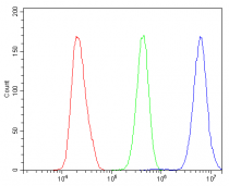
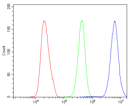
ARG41973 anti-PTBP2 antibody FACS image
Flow Cytometry: HeLa cells were blocked with 10% normal goat serum and then stained with ARG41973 anti-PTBP2 antibody (blue) at 1 µg/10^6 cells for 30 min at 20°C, followed by incubation with DyLight®488 labelled secondary antibody. Isotype control antibody (green) was Rabbit IgG (1 µg/10^6 cells) used under the same conditions. Unlabelled sample (red) was also used as a control.
-
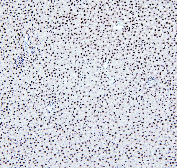
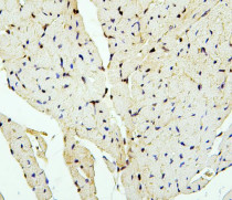
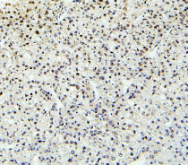
ARG41973 anti-PTBP2 antibody IHC-P image
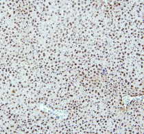
Immunohistochemistry: Paraffin-embedded Rat liver tissue. Antigen Retrieval: Heat mediation was performed in Citrate buffer (pH 6.0, epitope retrieval solution) for 20 min. The tissue section was blocked with 10% goat serum. The tissue section was then stained with ARG41973 anti-PTBP2 antibody at 1 µg/ml dilution, overnight at 4°C.
-
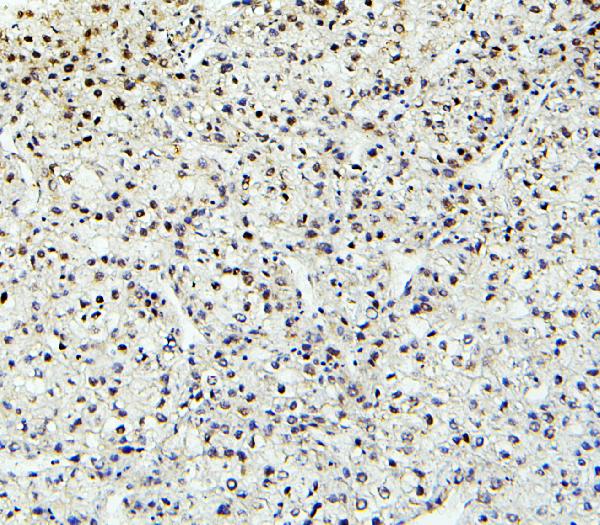
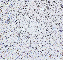
ARG41973 anti-PTBP2 antibody IHC-P image
Immunohistochemistry: Paraffin-embedded Human liver cancer tissue. Antigen Retrieval: Heat mediation was performed in Citrate buffer (pH 6.0, epitope retrieval solution) for 20 min. The tissue section was blocked with 10% goat serum. The tissue section was then stained with ARG41973 anti-PTBP2 antibody at 1 µg/ml dilution, overnight at 4°C.
-
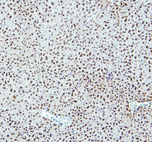
ARG41973 anti-PTBP2 antibody IHC-P image
Immunohistochemistry: Paraffin-embedded Mouse liver tissue. Antigen Retrieval: Heat mediation was performed in Citrate buffer (pH 6.0, epitope retrieval solution) for 20 min. The tissue section was blocked with 10% goat serum. The tissue section was then stained with ARG41973 anti-PTBP2 antibody at 1 µg/ml dilution, overnight at 4°C.
-
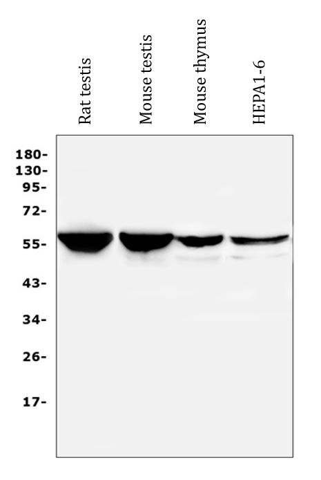
ARG41973 anti-PTBP2 antibody WB image
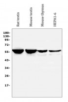
Western blot: 50 µg of samples under reducing conditions. Rat testis, Mouse testis, Mouse thymus and Mouse HEPA1-6 whole cell lysates stained with ARG41973 anti-PTBP2 antibody at 0.5 µg/ml dilution, overnight at 4°C.
-
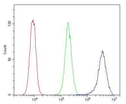
ARG41973 anti-PTBP2 antibody FACS image
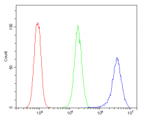
Flow Cytometry: THP-1 cells were blocked with 10% normal goat serum and then stained with ARG41973 anti-PTBP2 antibody (blue) at 1 µg/10^6 cells for 30 min at 20°C, followed by incubation with DyLight®488 labelled secondary antibody. Isotype control antibody (green) was Rabbit IgG (1 µg/10^6 cells) used under the same conditions. Unlabelled sample (red) was also used as a control.