ARG55445
anti-PSME2 antibody
anti-PSME2 antibody for Flow cytometry,IHC-Formalin-fixed paraffin-embedded sections,Western blot and Human,Mouse
Cell Biology and Cellular Response antibody
Overview
| Product Description | Rabbit Polyclonal antibody recognizes PSME2 |
|---|---|
| Tested Reactivity | Hu, Ms |
| Predict Reactivity | Bov, Rat |
| Tested Application | FACS, IHC-P, WB |
| Host | Rabbit |
| Clonality | Polyclonal |
| Isotype | IgG |
| Target Name | PSME2 |
| Antigen Species | Human |
| Immunogen | KLH-conjugated synthetic peptide corresponding to aa. 210-239 (C-terminus) of Human PSME2. |
| Conjugation | Un-conjugated |
| Alternate Names | 11S regulator complex subunit beta; REG-beta; PA28beta; Proteasome activator 28 subunit beta; Activator of multicatalytic protease subunit 2; PA28b; PA28B; Proteasome activator complex subunit 2; REGbeta |
Application Instructions
| Application Suggestion |
|
||||||||
|---|---|---|---|---|---|---|---|---|---|
| Application Note | * The dilutions indicate recommended starting dilutions and the optimal dilutions or concentrations should be determined by the scientist. | ||||||||
| Positive Control | HepG2 |
Properties
| Form | Liquid |
|---|---|
| Purification | This antibody is prepared by Saturated Ammonium Sulfate (SAS) precipitation followed by dialysis against PBS. |
| Buffer | PBS and 0.09% (W/V) Sodium azide |
| Preservative | 0.09% (W/V) Sodium azide |
| Storage Instruction | For continuous use, store undiluted antibody at 2-8°C for up to a week. For long-term storage, aliquot and store at -20°C or below. Storage in frost free freezers is not recommended. Avoid repeated freeze/thaw cycles. Suggest spin the vial prior to opening. The antibody solution should be gently mixed before use. |
| Note | For laboratory research only, not for drug, diagnostic or other use. |
Bioinformation
| Database Links |
Swiss-port # P97372 Mouse Proteasome activator complex subunit 2 Swiss-port # Q9UL46 Human Proteasome activator complex subunit 2 |
|---|---|
| Gene Symbol | PSME2 |
| Gene Full Name | proteasome (prosome, macropain) activator subunit 2 (PA28 beta) |
| Background | The 26S proteasome is a multicatalytic proteinase complex with a highly ordered structure composed of 2 complexes, a 20S core and a 19S regulator. The 20S core is composed of 4 rings of 28 non-identical subunits; 2 rings are composed of 7 alpha subunits and 2 rings are composed of 7 beta subunits. The 19S regulator is composed of a base, which contains 6 ATPase subunits and 2 non-ATPase subunits, and a lid, which contains up to 10 non-ATPase subunits. Proteasomes are distributed throughout eukaryotic cells at a high concentration and cleave peptides in an ATP/ubiquitin-dependent process in a non-lysosomal pathway. An essential function of a modified proteasome, the immunoproteasome, is the processing of class I MHC peptides. The immunoproteasome contains an alternate regulator, referred to as the 11S regulator or PA28, that replaces the 19S regulator. Three subunits (alpha, beta and gamma) of the 11S regulator have been identified. This gene encodes the beta subunit of the 11S regulator, one of the two 11S subunits that is induced by gamma-interferon. Three beta and three alpha subunits combine to form a heterohexameric ring. Six pseudogenes have been identified on chromosomes 4, 5, 8, 10 and 13. [provided by RefSeq, Jul 2008] |
| Function | Implicated in immunoproteasome assembly and required for efficient antigen processing. The PA28 activator complex enhances the generation of class I binding peptides by altering the cleavage pattern of the proteasome. [UniProt] |
| Research Area | Cell Biology and Cellular Response antibody |
| Calculated MW | 27 kDa |
Images (3) Click the Picture to Zoom In
-

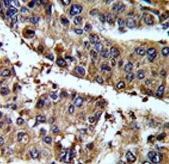
ARG55445 anti-PSME2 antibody IHC-P image
Immunohistochemistry: Formalin-fixed and paraffin-embedded Mouse hepatocarcinoma stained with ARG55445 anti-PSME2 antibody.
-
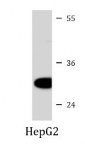
ARG55445 anti-PSME2 antibody WB image
Western blot: 35 µg of HepG2 cell lysate stained with ARG55445 anti-PSME2 antibody at 1:1000 dilution.
-
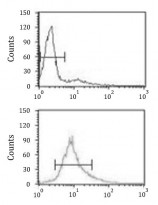
ARG55445 anti-PSME2 antibody FACS image
Flow Cytometry: HL-60 cells stained with ARG55445 anti-PSME2 antibody (bottom histogram) or without primary antibody control (top histogram), followed by incubation with FITC labelled secondary antibody.