ARG43990
anti-PSMA1 antibody
anti-PSMA1 antibody for ELISA,Flow cytometry,IHC-Formalin-fixed paraffin-embedded sections,Western blot and Human,Mouse,Rat
Overview
| Product Description | Rabbit Polyclonal antibody recognizes PSMA1 |
|---|---|
| Tested Reactivity | Hu, Ms, Rat |
| Tested Application | ELISA, FACS, IHC-P, WB |
| Host | Rabbit |
| Clonality | Polyclonal |
| Isotype | IgG |
| Target Name | PSMA1 |
| Antigen Species | Human |
| Immunogen | Human PSMA1 recombinant protein |
| Expression System | E.coli |
| Conjugation | Un-conjugated |
| Protein Full Name | Proteasome subunit alpha type-1 |
| Alternate Names | PSMA1; Proteasome 20S Subunit Alpha 1; PROS30; HC2; NU; Proteasome (Prosome, Macropain) Subunit, Alpha Type, 1; Multicatalytic Endopeptidase Complex Subunit C2; Proteasome Subunit Alpha Type-1; Proteasome Subunit Alpha 1; 30 KDa Prosomal Protein; Proteasome Component C2; Macropain Subunit C2; Proteasome Nu Chain; MGC14542; MGC14575; MGC14751; MGC21459; MGC22853; MGC23915; MGC1667; PROS-30; Epididymis Secretory Protein Li 275; Proteasome Subunit, Alpha-Type, 1; Testicular Tissue Protein Li 150; Proteasome Subunit Alpha 6; Proteasome Subunit Α6; Proteasome Subunit Nu; Macropain Subunit Nu; Protein P30-33K; EC 3.4.25.1 ; HEL-S-275; PSC2 |
Application Instructions
| Application Suggestion |
|
||||||||||
|---|---|---|---|---|---|---|---|---|---|---|---|
| Application Note | * The dilutions indicate recommended starting dilutions and the optimal dilutions or concentrations should be determined by the scientist. |
Properties
| Form | Liquid |
|---|---|
| Purification | Affinity purified with Immunogen. |
| Buffer | 0.9% NaCl, 0.2% Na2HPO4 and 4% Trehalose. |
| Stabilizer | 4% Trehalose |
| Concentration | 0.5 mg/ml |
| Storage Instruction | For continuous use, store undiluted antibody at 2-8°C for up to a week. For long-term storage, aliquot and store at -20°C or below. Storage in frost free freezers is not recommended. Avoid repeated freeze/thaw cycles. Suggest spin the vial prior to opening. The antibody solution should be gently mixed before use. |
| Note | For laboratory research only, not for drug, diagnostic or other use. |
Bioinformation
| Database Links | |
|---|---|
| Gene Symbol | PSMA1 |
| Gene Full Name | Proteasome 20S Subunit Alpha 1 |
| Background | he proteasome is a multicatalytic proteinase complex with a highly ordered ring-shaped 20S core structure. The core structure is composed of 4 rings of 28 non-identical subunits; 2 rings are composed of 7 alpha subunits and 2 rings are composed of 7 beta subunits. Proteasomes are distributed throughout eukaryotic cells at a high concentration and cleave peptides in an ATP/ubiquitin-dependent process in a non-lysosomal pathway. An essential function of a modified proteasome, the immunoproteasome, is the processing of class I MHC peptides. This gene encodes a member of the peptidase T1A family, that is a 20S core alpha subunit. Alternative splicing results in multiple transcript variants encoding distinct isoforms. |
| Function | Component of the 20S core proteasome complex involved in the proteolytic degradation of most intracellular proteins. This complex plays numerous essential roles within the cell by associating with different regulatory particles. Associated with two 19S regulatory particles, forms the 26S proteasome and thus participates in the ATP-dependent degradation of ubiquitinated proteins. The 26S proteasome plays a key role in the maintenance of protein homeostasis by removing misfolded or damaged proteins that could impair cellular functions, and by removing proteins whose functions are no longer required. Associated with the PA200 or PA28, the 20S proteasome mediates ubiquitin-independent protein degradation. This type of proteolysis is required in several pathways including spermatogenesis (20S-PA200 complex) or generation of a subset of MHC class I-presented antigenic peptides (20S-PA28 complex). |
| Cellular Localization | Cytoplasm, Nucleus, Proteasome |
| Calculated MW | 30 kDa |
| PTM | Acetylation, Glycoprotein, Isopeptide bond, Phosphoprotein, Ubl conjugation |
Images (8) Click the Picture to Zoom In
-
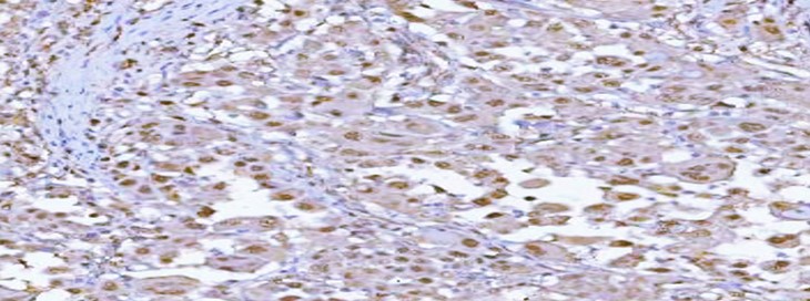
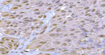
ARG43990 anti-PSMA1 antibody IHC-P image
Immunohistochemistry: Human lung adenocarcinoma stained with ARG43990 anti-PSMA1 antibody at 2 μg/ml dilution.
-
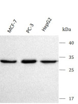
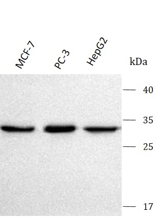
ARG43990 anti-PSMA1 antibody WB image
Western blot: MCF-7, PC-3 and HepG2 stained with ARG43990 anti-PSMA1 antibody at 0.5 μg/mL dilution.
-
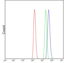
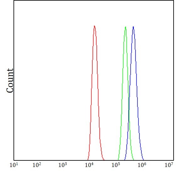
ARG43990 anti-PSMA1 antibody FACS image
Flow Cytometry: Raji cells stained with ARG43990 anti-PSMA1 antibody (blue) at 1 μg/1x10^6 cells dilution.
-
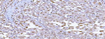
ARG43990 anti-PSMA1 antibody IHC-P image
Immunohistochemistry: Human urothelial carcinoma stained with ARG43990 anti-PSMA1 antibody at 2 μg/ml dilution.
-
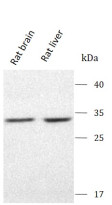
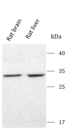
ARG43990 anti-PSMA1 antibody WB image
Western blot: Rat brain and Rat liver stained with ARG43990 anti-PSMA1 antibody at 0.5 μg/mL dilution.
-
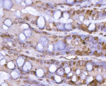
ARG43990 anti-PSMA1 antibody IHC-P image
Immunohistochemistry: Rat colon stained with ARG43990 anti-PSMA1 antibody at 2 μg/ml dilution.
-
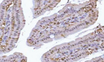
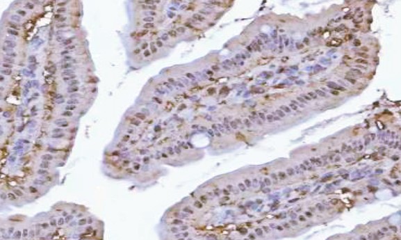
ARG43990 anti-PSMA1 antibody IHC-P image
Immunohistochemistry: Mouse colon stained with ARG43990 anti-PSMA1 antibody at 2 μg/ml dilution.
-

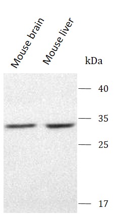
ARG43990 anti-PSMA1 antibody WB image
Western blot: Mouse brain and Mouse liver stained with ARG43990 anti-PSMA1 antibody at 0.5 μg/mL dilution.