ARG43986
anti-PSAP antibody
anti-PSAP antibody for ELISA,Flow cytometry,ICC/IF,IHC-Formalin-fixed paraffin-embedded sections,Western blot and Human
Overview
| Product Description | Rabbit Polyclonal antibody recognizes PSAP |
|---|---|
| Tested Reactivity | Hu |
| Tested Application | ELISA, FACS, ICC/IF, IHC-P, WB |
| Host | Rabbit |
| Clonality | Polyclonal |
| Isotype | IgG |
| Target Name | PSAP |
| Antigen Species | Human |
| Immunogen | Human PSAP recombinant protein |
| Expression System | E.coli |
| Conjugation | Un-conjugated |
| Protein Full Name | Prosaposin |
| Alternate Names | PSAP; Prosaposin; GLBA; SAP1; Sphingolipid Activator Protein-1; Sphingolipid Activator Protein-2; Proactivator Polypeptide; Saposin-A; Saposin-B; Saposin-C; Saposin-D; SAP2; Variant Gaucher Disease And Variant Metachromatic Leukodystrophy; PARK24; PSAPD |
Application Instructions
| Application Suggestion |
|
||||||||||||
|---|---|---|---|---|---|---|---|---|---|---|---|---|---|
| Application Note | * The dilutions indicate recommended starting dilutions and the optimal dilutions or concentrations should be determined by the scientist. |
Properties
| Form | Liquid |
|---|---|
| Purification | Affinity purified with Immunogen. |
| Buffer | 0.9% NaCl, 0.2% Na2HPO4 and 4% Trehalose. |
| Stabilizer | 4% Trehalose |
| Concentration | 0.5 mg/ml |
| Storage Instruction | For continuous use, store undiluted antibody at 2-8°C for up to a week. For long-term storage, aliquot and store at -20°C or below. Storage in frost free freezers is not recommended. Avoid repeated freeze/thaw cycles. Suggest spin the vial prior to opening. The antibody solution should be gently mixed before use. |
| Note | For laboratory research only, not for drug, diagnostic or other use. |
Bioinformation
| Database Links | |
|---|---|
| Gene Symbol | PSAP |
| Gene Full Name | Prosaposin |
| Background | This gene encodes a highly conserved preproprotein that is proteolytically processed to generate four main cleavage products including saposins A, B, C, and D. Each domain of the precursor protein is approximately 80 amino acid residues long with nearly identical placement of cysteine residues and glycosylation sites. Saposins A-D localize primarily to the lysosomal compartment where they facilitate the catabolism of glycosphingolipids with short oligosaccharide groups. The precursor protein exists both as a secretory protein and as an integral membrane protein and has neurotrophic activities. Mutations in this gene have been associated with Gaucher disease and metachromatic leukodystrophy. Alternative splicing results in multiple transcript variants, at least one of which encodes an isoform that is proteolytically processed. |
| Function | Saposin-A and saposin-C stimulate the hydrolysis of glucosylceramide by beta-glucosylceramidase (EC 3.2.1.45) and galactosylceramide by beta-galactosylceramidase (EC 3.2.1.46). Saposin-C apparently acts by combining with the enzyme and acidic lipid to form an activated complex, rather than by solubilizing the substrate. |
| Cellular Localization | Lysosome, Secreted |
| Calculated MW | 58 kDa |
| PTM | Disulfide bond, Glycoprotein |
Images (4) Click the Picture to Zoom In
-
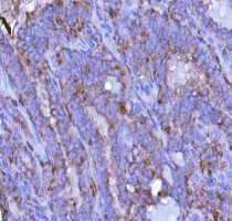
ARG43986 anti-PSAP antibody IHC-P image
Immunohistochemistry: Human thyroid papillary carcinoma stained with ARG43986 anti-PSAP antibody at 2 μg/ml dilution.
-
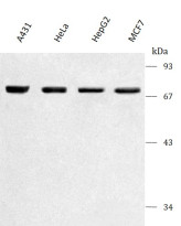
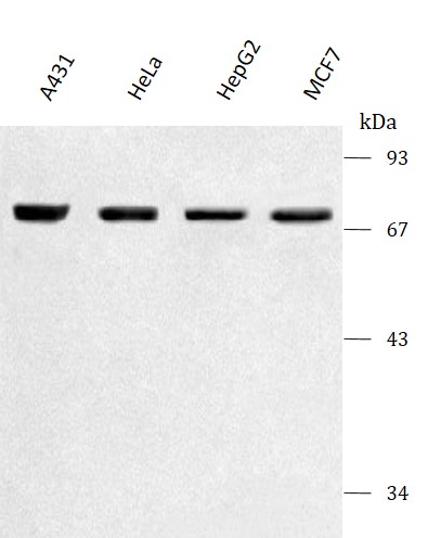
ARG43986 anti-PSAP antibody WB image
Western blot: A431, Hela, HepG2 and MCF-7 stained with ARG43986 anti-PSAP antibody at 0.5 μg/mL dilution.
-
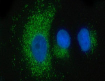
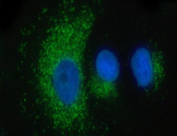
ARG43986 anti-PSAP antibody ICC/IF image
Immunofluorescence: A549 cells stained with ARG43986 anti-PSAP antibody at 5 μg/ml dilution.
-
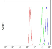
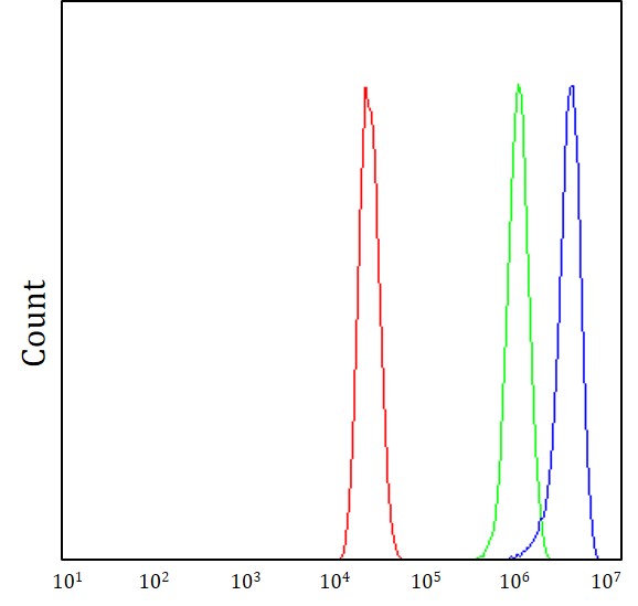
ARG43986 anti-PSAP antibody FACS image
Flow Cytometry: MCF-7 cells stained with ARG43986 anti-PSAP antibody (blue) at 1 μg/1x10^6 cells dilution.