ARG63153
anti-PPP1R15A / GADD34 antibody
anti-PPP1R15A / GADD34 antibody for Flow cytometry,ICC/IF,IHC-Formalin-fixed paraffin-embedded sections,Western blot and Human
Cell Biology and Cellular Response antibody; Gene Regulation antibody
Overview
| Product Description | Goat Polyclonal antibody recognizes PPP1R15A / GADD34 |
|---|---|
| Tested Reactivity | Hu |
| Tested Application | FACS, ICC/IF, IHC-P, WB |
| Host | Goat |
| Clonality | Polyclonal |
| Isotype | IgG |
| Target Name | PPP1R15A / GADD34 |
| Antigen Species | Human |
| Immunogen | C-AAALDLSGRRG |
| Conjugation | Un-conjugated |
| Alternate Names | Growth arrest and DNA damage-inducible protein GADD34; Protein phosphatase 1 regulatory subunit 15A; Myeloid differentiation primary response protein MyD116 homolog; GADD34 |
Application Instructions
| Application Suggestion |
|
||||||||||
|---|---|---|---|---|---|---|---|---|---|---|---|
| Application Note | WB: Recommend incubate at RT for 1h. IHC-P: Antigen Retrieval: Steam tissue section in Citrate buffer (pH 6.0). * The dilutions indicate recommended starting dilutions and the optimal dilutions or concentrations should be determined by the scientist. |
Properties
| Form | Liquid |
|---|---|
| Purification | Purified from goat serum by ammonium sulphate precipitation followed by antigen affinity chromatography using the immunizing peptide. |
| Buffer | Tris saline (pH 7.3), 0.02% Sodium azide and 0.5% BSA |
| Preservative | 0.02% Sodium azide |
| Stabilizer | 0.5% BSA |
| Concentration | 0.5 mg/ml |
| Storage Instruction | For continuous use, store undiluted antibody at 2-8°C for up to a week. For long-term storage, aliquot and store at -20°C or below. Storage in frost free freezers is not recommended. Avoid repeated freeze/thaw cycles. Suggest spin the vial prior to opening. The antibody solution should be gently mixed before use. |
| Note | For laboratory research only, not for drug, diagnostic or other use. |
Bioinformation
| Database Links |
Swiss-port # O75807 Human Protein phosphatase 1 regulatory subunit 15A |
|---|---|
| Background | This gene is a member of a group of genes whose transcript levels are increased following stressful growth arrest conditions and treatment with DNA-damaging agents. The induction of this gene by ionizing radiation occurs in certain cell lines regardless of p53 status, and its protein response is correlated with apoptosis following ionizing radiation. [provided by RefSeq, Jul 2008] |
| Research Area | Cell Biology and Cellular Response antibody; Gene Regulation antibody |
| Calculated MW | 73 kDa |
| PTM | Phosphorylated at multiple Ser/Thr residues. Phosphorylated on tyrosine by LYN; which impairs its antiproliferative activity. Phosphorylation at Tyr-262 enhances proteasomal degradation, this position is dephosphorylated by PTPN2. Polyubiquitinated. Exhibits a rapid proteasomal degradation with a half-life under 1 hour, ubiquitination depends on endoplasmic reticulum association. |
Images (5) Click the Picture to Zoom In
-

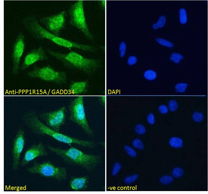
ARG63153 anti-PPP1R15A / GADD34 antibody ICC/IF image
Immunofluorescence: Paraformaldehyde fixed HeLa cells permeabilized with 0.15% Triton. Cells were stained with ARG63153 anti-PPP1R15A / GADD34 antibody (green) at 10 µg/ml dilution for 1 hour. DAPI (blue) for nuclear staining. Negative control: Unimmunized goat IgG (green) at 10 µg/ml dilution.
-
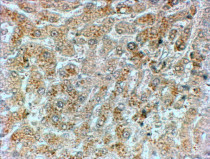
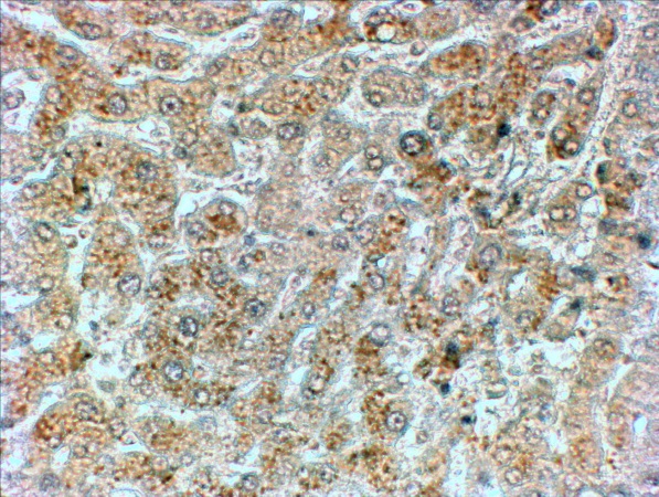
ARG63153 anti-PPP1R15A / GADD34 antibody IHC-P image
Immunohistochemistry: Paraffin-embedded Human liver tissue. Antigen Retrieval: Steam tissue section in Citrate buffer (pH 6.0). The tissue section was stained with ARG63153 anti-PPP1R15A / GADD34 antibody at 2 µg/ml dilution followed by HRP-staining.
-
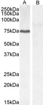
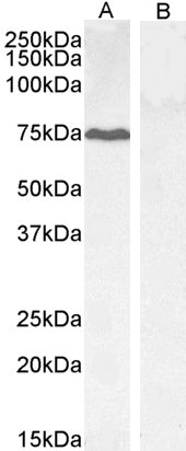
ARG63153 anti-PPP1R15A / GADD34 antibody WB image
Western blot: 35 µg of HepG2 (A) and KLY (B, negative control) cell lysates (in RIPA buffer) stained with ARG63153 anti-PPP1R15A / GADD34 antibody at 0.3 µg/ml dilution and incubated at RT for 1 hour.
-
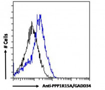
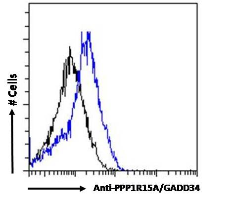
ARG63153 anti-PPP1R15A / GADD34 antibody FACS image
Flow Cytometry: Paraformaldehyde-fixed HepG2 cells permeabilized with 0.5% Triton. Cells were stained with ARG63153 anti-PPP1R15A / GADD34 antibody (blue line) at 10 µg/ml dilution for 1 hour, followed by incubation with Alexa FluorR 488 labelled secondary antibody. IgG control: Unimmunized goat IgG (black line).
-
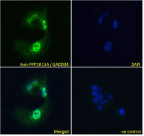
ARG63153 anti-PPP1R15A / GADD34 antibody ICC/IF image
Immunofluorescence: Paraformaldehyde fixed HepG2 cells permeabilized with 0.15% Triton. Cells were stained with ARG63153 anti-PPP1R15A / GADD34 antibody (green) at 10 µg/ml dilution for 1 hour. DAPI (blue) for nuclear staining. Negative control: Unimmunized goat IgG (green) at 10 µg/ml dilution.