ARG10651
anti-PLA2R antibody [12-6-5]
anti-PLA2R antibody [12-6-5] for Flow cytometry,ICC/IF,IHC-Formalin-fixed paraffin-embedded sections,Western blot and Human
Overview
| Product Description | Mouse Monoclonal antibody [12-6-5] recognizes PLA2R |
|---|---|
| Tested Reactivity | Hu |
| Tested Application | FACS, ICC/IF, IHC-P, WB |
| Host | Mouse |
| Clonality | Monoclonal |
| Clone | 12-6-5 |
| Isotype | IgG2b |
| Target Name | PLA2R |
| Antigen Species | Human |
| Immunogen | Recombinant protein around aa. 20-663 (extracellular sequence) of Human PLA2R. |
| Conjugation | Un-conjugated |
| Alternate Names | M-type receptor; Soluble PLA2-R; 180 kDa secretory phospholipase A2 receptor; Soluble PLA2R; PLA2G1R; CLEC13C; PLA2-R; C-type lectin domain family 13 member C; PLA2R; Secretory phospholipase A2 receptor; PLA2IR |
Application Instructions
| Application Suggestion |
|
||||||||||
|---|---|---|---|---|---|---|---|---|---|---|---|
| Application Note | IHC-P: Antigen Retrieval: 64 min heat in Cell Conditioner 1 (Tris-EDTA, pH 8.0 buffer). * The dilutions indicate recommended starting dilutions and the optimal dilutions or concentrations should be determined by the scientist. |
||||||||||
| Positive Control | Human normal kidney |
Properties
| Form | Liquid |
|---|---|
| Purification | Purification with Protein A. |
| Buffer | PBS and 0.02% Sodium azide. |
| Preservative | 0.02% Sodium azide |
| Storage Instruction | For continuous use, store undiluted antibody at 2-8°C for up to a week. For long-term storage, aliquot and store at -20°C. Storage in frost free freezers is not recommended. Avoid repeated freeze/thaw cycles. Suggest spin the vial prior to opening. The antibody solution should be gently mixed before use. |
| Note | For laboratory research only, not for drug, diagnostic or other use. |
Bioinformation
| Database Links |
Swiss-port # Q13018 Human Secretory phospholipase A2 receptor |
|---|---|
| Gene Symbol | PLA2R1 |
| Gene Full Name | phospholipase A2 receptor 1, 180kDa |
| Background | This gene represents a phospholipase A2 receptor. The encoded protein likely exists as both a transmembrane form and a soluble form. The transmembrane receptor may play a role in clearance of phospholipase A2, thereby inhibiting its action. Polymorphisms at this locus have been associated with susceptibility to idiopathic membranous nephropathy. Alternatively spliced transcript variants encoding different isoforms have been identified.[provided by RefSeq, Sep 2010] |
| Function | Receptor for secretory phospholipase A2 (sPLA2). Acts as a receptor for phosholipase sPLA2-IB/PLA2G1B but not sPLA2-IIA/PLA2G2A. Also able to bind to snake PA2-like toxins. Although its precise function remains unclear, binding of sPLA2 to its receptor participates in both positive and negative regulation of sPLA2 functions as well as clearance of sPLA2. Binding of sPLA2-IB/PLA2G1B induces various effects depending on the cell type, such as activation of the mitogen-activated protein kinase (MAPK) cascade to induce cell proliferation, the production of lipid mediators, selective release of arachidonic acid in bone marrow-derived mast cells. In neutrophils, binding of sPLA2-IB/PLA2G1B can activate p38 MAPK to stimulate elastase release and cell adhesion. May be involved in responses in proinflammatory cytokine productions during endotoxic shock. Also has endocytic properties and rapidly internalizes sPLA2 ligands, which is particularly important for the clearance of extracellular sPLA2s to protect their potent enzymatic activities. The soluble secretory phospholipase A2 receptor form is circulating and acts as a negative regulator of sPLA2 functions by blocking the biological functions of sPLA2-IB/PLA2G1B. [UniProt] |
| Calculated MW | 169 kDa |
| PTM | The secretory phospholipase A2 receptor form may be produced by the action of metalloproteinases. It contains all extracellular domains and only lacks transmembrane and cytosolic regions. It is however unclear whether this form is produced by proteolytic cleavage as suggested by some experiments, or by alternative splicing, as in the case of isoform 2 that shares all characteristics of secretory phospholipase A2 receptor form (By similarity). |
Images (6) Click the Picture to Zoom In
-
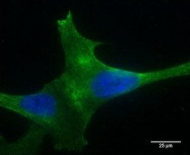
ARG10651 anti-PLA2R antibody [12-6-5] ICC/IF image
Immunofluorescence: PLA2R over-expressing podocytes revealing cell surface expression of the PLA2R receptor using ARG10651 anti-PLA2R antibody [12-6-5] at 1:400 dilution (40x lens).
-
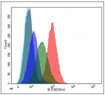
ARG10651 anti-PLA2R antibody [12-6-5] FACS image
Flow Cytometry: wild type (dark blue line) and over-expressing PLA2R podocytes (low expressor, green line; high expressor, red line) stained with ARG10651 anti-PLA2R antibody [12-6-5] at 1:200 dilution. Negative controls were unlabelled cells (turquoise line) and mouse IgG (grey line).
-
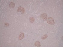
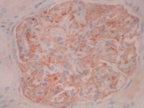
ARG10651 anti-PLA2R antibody [12-6-5] IHC-P image
Immunohistochemistry: Paraffin-embedded Normal Human kidney showing glomerular specific staining.
IHC Protocol Info: HIER pH8 (CC1) for 64 minutes, 36 minute ARG10651 anti-PLA2R antibody [12-6-5] incubation at RT at 1:5000 dilution. -
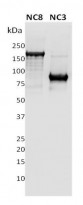
ARG10651 anti-PLA2R antibody [12-6-5] WB image
Western blot: 1 µg of recombinant full length extracellular domain of Human PLA2R (NC8) and a N-terminus fragment of PLA2R (NC3) were subjected to SDS PAGE followed by WB stained with ARG10651 anti-PLA2R antibody [12-6-5] at 1:10000 dilution.
-
ARG10651 anti-PLA2R antibody [12-6-5] IHC-P image
Immunohistochemistry: Paraffin-embedded Normal Human kidney showing podocyte specific staining.
IHC Protocol Info: HIER pH8 (CC1) for 64 minutes, 36 minute ARG10651 anti-PLA2R antibody [12-6-5] incubation at RT at 1:5000 dilution. -
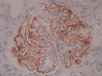
ARG10651 anti-PLA2R antibody [12-6-5] IHC-P image
Immunohistochemistry: Paraffin-embedded Human Membranous nephropathy kidney showing capillary wall of the glomerular basement membrane specific staining.
IHC Protocol Info: HIER pH8 (CC1) for 64 minutes, 36 minute ARG10651 anti-PLA2R antibody [12-6-5] incubation at RT at 1:5000 dilution.


