ARG65025
anti-PKD2 / Polycystin 2 antibody
anti-PKD2 / Polycystin 2 antibody for Western blot and Human
Metabolism antibody; Signaling Transduction antibody
Overview
| Product Description | Goat Polyclonal antibody recognizes PKD2 / Polycystin 2 |
|---|---|
| Tested Reactivity | Hu |
| Predict Reactivity | Ms, Rat, Cow |
| Tested Application | WB |
| Host | Goat |
| Clonality | Polyclonal |
| Isotype | IgG |
| Target Name | PKD2 / Polycystin 2 |
| Antigen Species | Human |
| Immunogen | C-ERAKLKRREVLGR |
| Conjugation | Un-conjugated |
| Alternate Names | Polycystwin; APKD2; Autosomal dominant polycystic kidney disease type II protein; PC2; Pc-2; Polycystic kidney disease 2 protein; R48321; TRPP2; Polycystin-2; PKD4; Transient receptor potential cation channel subfamily P member 2 |
Application Instructions
| Application Suggestion |
|
||||
|---|---|---|---|---|---|
| Application Note | WB: Recommend incubate at RT for 1h. * The dilutions indicate recommended starting dilutions and the optimal dilutions or concentrations should be determined by the scientist. |
Properties
| Form | Liquid |
|---|---|
| Purification | Purified from goat serum by ammonium sulphate precipitation followed by antigen affinity chromatography using the immunizing peptide. |
| Buffer | Tris saline (pH 7.3), 0.02% Sodium azide and 0.5% BSA |
| Preservative | 0.02% Sodium azide |
| Stabilizer | 0.5% BSA |
| Concentration | 0.5 mg/ml |
| Storage Instruction | For continuous use, store undiluted antibody at 2-8°C for up to a week. For long-term storage, aliquot and store at -20°C or below. Storage in frost free freezers is not recommended. Avoid repeated freeze/thaw cycles. Suggest spin the vial prior to opening. The antibody solution should be gently mixed before use. |
| Note | For laboratory research only, not for drug, diagnostic or other use. |
Bioinformation
| Database Links | |
|---|---|
| Gene Symbol | PKD2 |
| Gene Full Name | polycystin 2, transient receptor potential cation channel |
| Background | This gene encodes a member of the polycystin protein family. The encoded protein is a multi-pass membrane protein that functions as a calcium permeable cation channel, and is involved in calcium transport and calcium signaling in renal epithelial cells. This protein interacts with polycystin 1, and they may be partners in a common signaling cascade involved in tubular morphogenesis. Mutations in this gene are associated with autosomal dominant polycystic kidney disease type 2. [provided by RefSeq, Mar 2011] |
| Function | Component of a heteromeric calcium-permeable ion channel formed by PKD1 and PKD2 that is activated by interaction between PKD1 and a Wnt family member, such as WNT3A and WNT9B (PubMed:27214281). Can also form a functional, homotetrameric ion channel (PubMed:29899465). Functions as a cation channel involved in fluid-flow mechanosensation by the primary cilium in renal epithelium (PubMed:18695040). Functions as outward-rectifying K+ channel, but is also permeable to Ca2+, and to a much lesser degree also to Na+ (PubMed:11854751, PubMed:15692563, PubMed:27071085, PubMed:27991905). May contribute to the release of Ca2+ stores from the endoplasmic reticulum (PubMed:11854751, PubMed:20881056). Together with TRPV4, forms mechano- and thermosensitive channels in cilium (PubMed:18695040). PKD1 and PKD2 may function through a common signaling pathway that is necessary to maintain the normal, differentiated state of renal tubule cells. Acts as a regulator of cilium length, together with PKD1. The dynamic control of cilium length is essential in the regulation of mechanotransductive signaling. The cilium length response creates a negative feedback loop whereby fluid shear-mediated deflection of the primary cilium, which decreases intracellular cAMP, leads to cilium shortening and thus decreases flow-induced signaling. Also involved in left-right axis specification via its role in sensing nodal flow; forms a complex with PKD1L1 in cilia to facilitate flow detection in left-right patterning. Detection of asymmetric nodal flow gives rise to a Ca2+ signal that is required for normal, asymmetric expression of genes involved in the specification of body left-right laterality. [UniProt] |
| Research Area | Metabolism antibody; Signaling Transduction antibody |
| Calculated MW | 110 kDa |
| PTM | Phosphorylated. Phosphorylation is important for protein function; a mutant that lacks the N-terminal phosphorylation sites cannot complement a zebrafish pkd2-deficient mutant (PubMed:16551655). PKD-mediated phosphorylation at the C-terminus regulates its function in the release of Ca(2+) stores from the endoplasmic reticulum (PubMed:20881056). PKA-mediated phosphorylation at a C-terminal site strongly increases the open probability of the channel, but does not increase single channel conductance (PubMed:26269590). N-glycosylated. The four subunits in a tetramer probably differ in the extent of glycosylation; simultaneous glycosylation of all experimentally validated sites would probably create steric hindrance. Thus, glycosylation at Asn-305 is not compatible with glycosylation at Asn-328; only one of these two residues is glycosylated at a given time. |
Images (1) Click the Picture to Zoom In
-

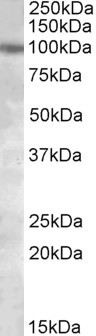
ARG65025 anti-PKD2 / Polycystin 2 antibody WB image
Western blot: 35 µg of HeLa cell lysate (in RIPA buffer) stained with ARG65025 anti-PKD2 / Polycystin 2 antibody at 1 µg/ml dilution and incubated at RT for 1 hour.