ARG63087
anti-PKA C alpha antibody [6D2.1]
anti-PKA C alpha antibody [6D2.1] for Western blot and Human
Cancer antibody; Metabolism antibody; Signaling Transduction antibody
Overview
| Product Description | Mouse Monoclonal antibody [6D2.1] recognizes PKA C alpha |
|---|---|
| Tested Reactivity | Hu |
| Predict Reactivity | Dog, Pig, Sheep |
| Tested Application | WB |
| Specificity | The clone 6D2.1 strongly reacts with human proteinkinase A catalytic (PKAc) alpha subunit, and weakly with PKAc gamma subunit (both around 40 kDa). The recognized epitope of PKAc alpha is identical between man, sheep, pig, ox and dog. |
| Host | Mouse |
| Clonality | Monoclonal |
| Clone | 6D2.1 |
| Isotype | IgG1 |
| Target Name | PKA C alpha |
| Antigen Species | Human |
| Immunogen | Peptide corresponding to amino acids ESPAQNTAHLDQFERIK of human proteinkinase A c alpha (PKAc alpha). |
| Conjugation | Un-conjugated |
| Alternate Names | EC 2.7.11.11; PKA C-alpha; PKACA; cAMP-dependent protein kinase catalytic subunit alpha; PPNAD4 |
Application Instructions
| Application Suggestion |
|
||||
|---|---|---|---|---|---|
| Application Note | WB: To detect PKAc gamma, use a more concentrated lysate from a tissue expressing this subunit (testis). * The dilutions indicate recommended starting dilutions and the optimal dilutions or concentrations should be determined by the scientist. |
||||
| Positive Control | HeLa |
Properties
| Form | Liquid |
|---|---|
| Purification | Purified from cell culture supernatant by protein-A affinity chromatography. |
| Purity | > 95% (by SDS-PAGE) |
| Buffer | PBS (pH 7.4) and 15 mM Sodium azide |
| Preservative | 15 mM Sodium azide |
| Concentration | 1 mg/ml |
| Storage Instruction | For continuous use, store undiluted antibody at 2-8°C for up to a week. For long-term storage, aliquot and store at -20°C or below. Storage in frost free freezers is not recommended. Avoid repeated freeze/thaw cycles. Suggest spin the vial prior to opening. The antibody solution should be gently mixed before use. |
| Note | For laboratory research only, not for drug, diagnostic or other use. |
Bioinformation
| Database Links |
Swiss-port # P17612 Human cAMP-dependent protein kinase catalytic subunit alpha |
|---|---|
| Gene Symbol | PRKACA |
| Gene Full Name | protein kinase, cAMP-dependent, catalytic, alpha |
| Background | Protein kinase A (PKA, cAMP-dependent protein kinase) is a key element of a ubiquitous signaling pathway important in the cell cycle, cellular communication, memory formation and behavior. PKA is composed of two catalytic (PKAc; Protein Kinase A catalytic subunit) and two regulatory subunits (PKAr). Upon binding cAMP, the complex dissociates to PKAr dimer and two activated PKAc ser/thr protein kinase catalytic monomers. The released PKAc can translocate into the nucleus and exert a regulatory role in the activation of multiple nuclear hormone receptors. However, PKAc-mediated activation of tonicity-dependent gene expression is cAMP independent. Humans express three types of PKAc subunit – PKAc alpha is present in most human tissues, PKAc beta and gamma are tissue-specific, the later is found in testes. |
| Function | Phosphorylates a large number of substrates in the cytoplasm and the nucleus. Regulates the abundance of compartmentalized pools of its regulatory subunits through phosphorylation of PJA2 which binds and ubiquitinates these subunits, leading to their subsequent proteolysis. Phosphorylates CDC25B, ABL1, NFKB1, CLDN3, PSMC5/RPT6, PJA2, RYR2, RORA and VASP. RORA is activated by phosphorylation. Required for glucose-mediated adipogenic differentiation increase and osteogenic differentiation inhibition from osteoblasts. Involved in the regulation of platelets in response to thrombin and collagen; maintains circulating platelets in a resting state by phosphorylating proteins in numerous platelet inhibitory pathways when in complex with NF-kappa-B (NFKB1 and NFKB2) and I-kappa-B-alpha (NFKBIA), but thrombin and collagen disrupt these complexes and free active PRKACA stimulates platelets and leads to platelet aggregation by phosphorylating VASP. Prevents the antiproliferative and anti-invasive effects of alpha-difluoromethylornithine in breast cancer cells when activated. RYR2 channel activity is potentiated by phosphorylation in presence of luminal Ca(2+), leading to reduced amplitude and increased frequency of store overload-induced Ca(2+) release (SOICR) characterized by an increased rate of Ca(2+) release and propagation velocity of spontaneous Ca(2+) waves, despite reduced wave amplitude and resting cytosolic Ca(2+). PSMC5/RPT6 activation by phosphorylation stimulates proteasome. Negatively regulates tight junctions (TJs) in ovarian cancer cells via CLDN3 phosphorylation. NFKB1 phosphorylation promotes NF-kappa-B p50-p50 DNA binding. Involved in embryonic development by down-regulating the Hedgehog (Hh) signaling pathway that determines embryo pattern formation and morphogenesis. Prevents meiosis resumption in prophase-arrested oocytes via CDC25B inactivation by phosphorylation. May also regulate rapid eye movement (REM) sleep in the pedunculopontine tegmental (PPT). Phosphorylates APOBEC3G and AICDA. Isoform 2 phosphorylates and activates ABL1 in sperm flagellum to promote spermatozoa capacitation. [UniProt] |
| Research Area | Cancer antibody; Metabolism antibody; Signaling Transduction antibody |
| Calculated MW | 41 kDa |
| PTM | Asn-3 is partially deaminated to Asp giving rise to 2 major isoelectric variants, called CB and CA respectively. Autophosphorylated. Phosphorylation is enhanced by vitamin K(2). Phosphorylated on threonine and serine residues. Phosphorylation on Thr-198 is required for full activity. Phosphorylated at Tyr-331 by activated receptor tyrosine kinases EGFR and PDGFR; this increases catalytic efficienncy. |
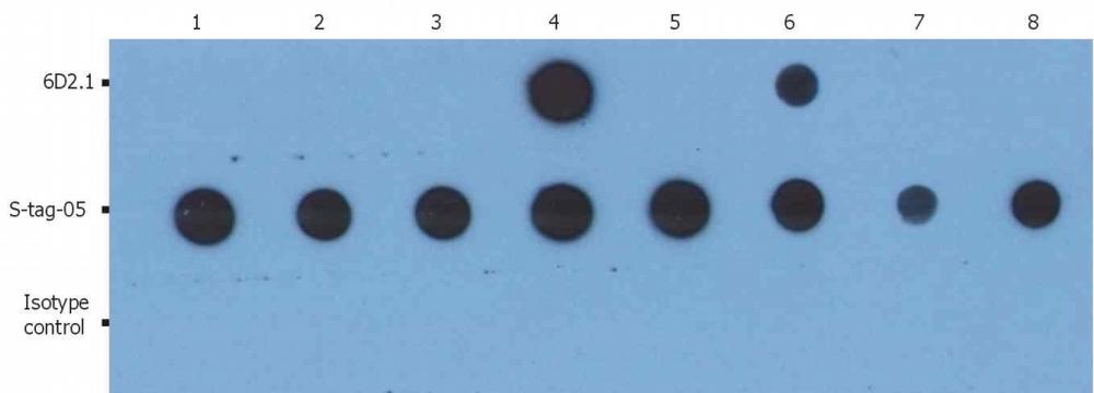
Images (1) Click the Picture to Zoom In
-

ARG63087 anti-PKAc antibody [6D2.1] Dot Blot image
Dot Blot: The total amount of material spotted on the nitrocellulose membrane is 5 ng/spot. (1): GST-Akt1 Lane (2): GST-Akt2 (3): GST-Akt3 (4): GST-PKAc alpha (5): GST-PKAc beta (6): GST-PKAc gamma (7): GST-MEK 1 (8): GST stained with ARG63087 anti-PKAc antibody [6D2.1].