ARG22358
anti-PINK1 antibody [S4-15]
anti-PINK1 antibody [S4-15] for ICC/IF,Western blot and Human,Rat
Overview
| Product Description | Mouse Monoclonal antibody [S4-15] recognizes PINK1 |
|---|---|
| Tested Reactivity | Hu, Rat |
| Predict Reactivity | Ms |
| Tested Application | ICC/IF, WB |
| Host | Mouse |
| Clonality | Monoclonal |
| Clone | S4-15 |
| Isotype | IgG1 |
| Target Name | PINK1 |
| Antigen Species | Human |
| Immunogen | Fusion protein around aa. 112-496 (cytoplasmic C-terminus) of Human PINK1. 82% identical to Rat and 81% identical to Mouse. > 30% identity with DMPK. |
| Conjugation | Un-conjugated |
| Alternate Names | PARK6; BRPK; PTEN-induced putative kinase protein 1; Serine/threonine-protein kinase PINK1, mitochondrial; EC 2.7.11.1 |
Application Instructions
| Application Suggestion |
|
||||||
|---|---|---|---|---|---|---|---|
| Application Note | * The dilutions indicate recommended starting dilutions and the optimal dilutions or concentrations should be determined by the scientist. |
Properties
| Form | Liquid |
|---|---|
| Purification | Purification with Protein G. |
| Buffer | PBS (pH 7.4), 0.1% Sodium azide and 50% Glycerol. |
| Preservative | 0.1% Sodium azide |
| Stabilizer | 50% Glycerol |
| Concentration | 1 mg/ml |
| Storage Instruction | For continuous use, store undiluted antibody at 2-8°C for up to a week. For long-term storage, aliquot and store at -20°C. Storage in frost free freezers is not recommended. Avoid repeated freeze/thaw cycles. Suggest spin the vial prior to opening. The antibody solution should be gently mixed before use. |
| Note | For laboratory research only, not for drug, diagnostic or other use. |
Bioinformation
| Database Links |
Swiss-port # Q9BXM7 Human Serine/threonine-protein kinase PINK1, mitochondrial |
|---|---|
| Gene Symbol | PINK1 |
| Gene Full Name | PTEN induced putative kinase 1 |
| Background | This gene encodes a serine/threonine protein kinase that localizes to mitochondria. It is thought to protect cells from stress-induced mitochondrial dysfunction. Mutations in this gene cause one form of autosomal recessive early-onset Parkinson disease. [provided by RefSeq, Jul 2008] |
| Function | Protects against mitochondrial dysfunction during cellular stress by phosphorylating mitochondrial proteins. Involved in the clearance of damaged mitochondria via selective autophagy (mitophagy) by mediating activation and translocation of PARK2. Targets PARK2 to dysfunctional depolarized mitochondria through the phosphorylation of MFN2. Activates PARK2 in 2 steps: (1) by mediating phosphorylation at 'Ser-65' of PARK2 and (2) mediating phosphorylation of ubiquitin, converting PARK2 to its fully-active form. [UniProt] |
| Highlight | Related products: PINK1 antibodies; Anti-Mouse IgG secondary antibodies; Related news: Astrocyte-to-neuron conversion for Parkinson's disease treatment |
| Calculated MW | 63 kDa |
| PTM | Autophosphorylation at Ser-228 and Ser-402 is essential for Parkin/PRKN recruitment to depolarized mitochondria. Two shorter forms of 55 kDa and 48 kDa seem to be produced by proteolytic cleavage and localize mainly in cytosol. |
Images (2) Click the Picture to Zoom In
-
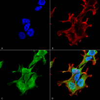
ARG22358 anti-PINK1 antibody [S4-15] ICC/IF image
Immunocytochemistry: Human Neuroblastoma cell line SK-N-BE. Fixation: 4% Formaldehyde for 15 min at RT. Cells were stained with ARG22358 anti-PINK1 antibody [S4-15] at 1:100 dilution (60 min, RT). Magnification: 60X. (A) DAPI (blue) nuclear stain, (B) Phalloidin Texas Red F-Actin stain, (C) ARG22358 anti-PINK1 antibody [S4-15] (green), and (D) Composite.
-
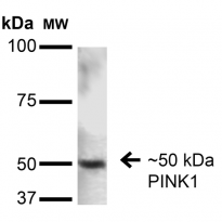
ARG22358 anti-PINK1 antibody [S4-15] WB image
Western blot: 15 µg of Rat Brain stained with ARG22358 anti-PINK1 antibody [S4-15] at 1:200 dilution (16 hours, 4°C). Block: 2% BSA and 2% Skim Milk in 1X TBST.


