ARG64304
anti-PINK1 antibody
anti-PINK1 antibody for Flow cytometry,Western blot and Rat
Cell Biology and Cellular Response antibody; Metabolism antibody; Neuroscience antibody; Signaling Transduction antibody
Overview
| Product Description | Goat Polyclonal antibody recognizes PINK1 |
|---|---|
| Tested Reactivity | Rat |
| Predict Reactivity | Hu |
| Tested Application | FACS, WB |
| Host | Goat |
| Clonality | Polyclonal |
| Isotype | IgG |
| Target Name | PINK1 |
| Antigen Species | Human |
| Immunogen | C-QGKAHLESRSYQEAQ |
| Conjugation | Un-conjugated |
| Alternate Names | PARK6; BRPK; PTEN-induced putative kinase protein 1; Serine/threonine-protein kinase PINK1, mitochondrial; EC 2.7.11.1 |
Application Instructions
| Application Suggestion |
|
||||||
|---|---|---|---|---|---|---|---|
| Application Note | WB: Recommend incubate at RT for 1h. * The dilutions indicate recommended starting dilutions and the optimal dilutions or concentrations should be determined by the scientist. |
Properties
| Form | Liquid |
|---|---|
| Purification | Purified from goat serum by ammonium sulphate precipitation followed by antigen affinity chromatography using the immunizing peptide. |
| Buffer | Tris saline (pH 7.3), 0.02% Sodium azide and 0.5% BSA |
| Preservative | 0.02% Sodium azide |
| Stabilizer | 0.5% BSA |
| Concentration | 0.5 mg/ml |
| Storage Instruction | For continuous use, store undiluted antibody at 2-8°C for up to a week. For long-term storage, aliquot and store at -20°C or below. Storage in frost free freezers is not recommended. Avoid repeated freeze/thaw cycles. Suggest spin the vial prior to opening. The antibody solution should be gently mixed before use. |
| Note | For laboratory research only, not for drug, diagnostic or other use. |
Bioinformation
| Background | This gene encodes a serine/threonine protein kinase that localizes to mitochondria. It is thought to protect cells from stress-induced mitochondrial dysfunction. Mutations in this gene cause one form of autosomal recessive early-onset Parkinson disease. [provided by RefSeq, Jul 2008] |
|---|---|
| Highlight | Related products: PINK1 antibodies; Anti-Goat IgG secondary antibodies; Related news: Astrocyte-to-neuron conversion for Parkinson's disease treatment |
| Research Area | Cell Biology and Cellular Response antibody; Metabolism antibody; Neuroscience antibody; Signaling Transduction antibody |
| Calculated MW | 63 kDa |
| PTM | Autophosphorylation at Ser-228 and Ser-402 is essential for Parkin/PRKN recruitment to depolarized mitochondria. Two shorter forms of 55 kDa and 48 kDa seem to be produced by proteolytic cleavage and localize mainly in cytosol. |
Images (2) Click the Picture to Zoom In
-

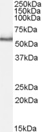
ARG64304 anti-PINK1 antibody WB image
Western Blot: Rat Testis lysate (35 µg protein in RIPA buffer) stained with ARG64304 anti-PINK1 antibody at 1 µg/ml dilution.
-
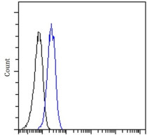
ARG64304 anti-PINK1 antibody FACS image
Flow Cytometry: Jurkat stained with ARG64304 anti-PINK1 antibody at 10 µg/ml dilution.