ARG58149
anti-PIM1 antibody
anti-PIM1 antibody for ICC/IF,IHC-Formalin-fixed paraffin-embedded sections,Western blot and Human,Mouse
Overview
| Product Description | Rabbit Polyclonal antibody recognizes PIM1 |
|---|---|
| Tested Reactivity | Hu, Ms |
| Tested Application | ICC/IF, IHC-P, WB |
| Host | Rabbit |
| Clonality | Polyclonal |
| Isotype | IgG |
| Target Name | PIM1 |
| Antigen Species | Human |
| Immunogen | Synthetic peptide derived from Human PIM1. |
| Conjugation | Un-conjugated |
| Alternate Names | EC 2.7.11.1; PIM; Serine/threonine-protein kinase pim-1 |
Application Instructions
| Application Suggestion |
|
||||||||
|---|---|---|---|---|---|---|---|---|---|
| Application Note | * The dilutions indicate recommended starting dilutions and the optimal dilutions or concentrations should be determined by the scientist. | ||||||||
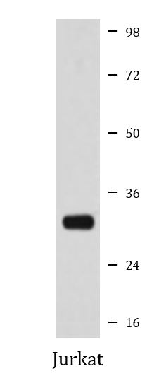
| Positive Control | Jurkat | ||||||||
| Observed Size | ~ 32 kDa |
Properties
| Form | Liquid |
|---|---|
| Purification | Affinity purified. |
| Buffer | PBS (pH 7.4), 0.02% Sodium azide and 50% Glycerol. |
| Preservative | 0.02% Sodium azide |
| Stabilizer | 50% Glycerol |
| Storage Instruction | For continuous use, store undiluted antibody at 2-8°C for up to a week. For long-term storage, aliquot and store at -20°C. Storage in frost free freezers is not recommended. Avoid repeated freeze/thaw cycles. Suggest spin the vial prior to opening. The antibody solution should be gently mixed before use. |
| Note | For laboratory research only, not for drug, diagnostic or other use. |
Bioinformation
| Database Links |
Swiss-port # P11309 Human Serine/threonine-protein kinase pim-1 |
|---|---|
| Gene Symbol | PIM1 |
| Gene Full Name | Pim-1 proto-oncogene, serine/threonine kinase |
| Background | The protein encoded by this gene belongs to the Ser/Thr protein kinase family, and PIM subfamily. This gene is expressed primarily in B-lymphoid and myeloid cell lines, and is overexpressed in hematopoietic malignancies and in prostate cancer. It plays a role in signal transduction in blood cells, contributing to both cell proliferation and survival, and thus provides a selective advantage in tumorigenesis. Both the human and orthologous mouse genes have been reported to encode two isoforms (with preferential cellular localization) resulting from the use of alternative in-frame translation initiation codons, the upstream non-AUG (CUG) and downstream AUG codons (PMIDs:16186805, 1825810).[provided by RefSeq, Aug 2011] |
| Function | Proto-oncogene with serine/threonine kinase activity involved in cell survival and cell proliferation and thus providing a selective advantage in tumorigenesis. Exerts its oncogenic activity through: the regulation of MYC transcriptional activity, the regulation of cell cycle progression and by phosphorylation and inhibition of proapoptotic proteins (BAD, MAP3K5, FOXO3). Phosphorylation of MYC leads to an increase of MYC protein stability and thereby an increase of transcriptional activity. The stabilization of MYC exerted by PIM1 might explain partly the strong synergism between these two oncogenes in tumorigenesis. Mediates survival signaling through phosphorylation of BAD, which induces release of the anti-apoptotic protein Bcl-X(L)/BCL2L1. Phosphorylation of MAP3K5, an other proapoptotic protein, by PIM1, significantly decreases MAP3K5 kinase activity and inhibits MAP3K5-mediated phosphorylation of JNK and JNK/p38MAPK subsequently reducing caspase-3 activation and cell apoptosis. Stimulates cell cycle progression at the G1-S and G2-M transitions by phosphorylation of CDC25A and CDC25C. Phosphorylation of CDKN1A, a regulator of cell cycle progression at G1, results in the relocation of CDKN1A to the cytoplasm and enhanced CDKN1A protein stability. Promote cell cycle progression and tumorigenesis by down-regulating expression of a regulator of cell cycle progression, CDKN1B, at both transcriptional and post-translational levels. Phosphorylation of CDKN1B,induces 14-3-3-proteins binding, nuclear export and proteasome-dependent degradation. May affect the structure or silencing of chromatin by phosphorylating HP1 gamma/CBX3. Acts also as a regulator of homing and migration of bone marrow cells involving functional interaction with the CXCL12-CXCR4 signaling axis. [UniProt] |
| Cellular Localization | Cell membrane. [UniProt] |
| Calculated MW | 36 kDa |
| PTM | Autophosphorylated on both serine/threonine and tyrosine residues. Phosphorylated. Interaction with PPP2CA promotes dephosphorylation. Ubiquitinated, leading to proteasomal degradation. [UniProt] |
Images (2) Click the Picture to Zoom In
-

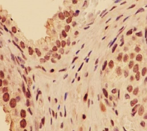
ARG58149 anti-PIM1 antibody IHC-P image
Immunohistochemistry: Paraffin-embedded Human prostate carcinoma stained with ARG58149 anti-PIM1 antibody.
-
ARG58149 anti-PIM1 antibody WB image
Western blot: Jurkat cell lysate stained with ARG58149 anti-PIM1 antibody.