ARG20571
anti-PI3 Kinase p85 alpha antibody [M253]
anti-PI3 Kinase p85 alpha antibody [M253] for ICC/IF,IHC-Formalin-fixed paraffin-embedded sections,Western blot and Human
Cancer antibody; Immune System antibody; Signaling Transduction antibody
Overview
| Product Description | Mouse Monoclonal antibody [M253] recognizes PI3 Kinase p85 alpha |
|---|---|
| Tested Reactivity | Hu |
| Predict Reactivity | Ms, Rat, Chk |
| Tested Application | ICC/IF, IHC-P, WB |
| Specificity | This antibody detects an 85 kDa band corresponding to the p85 subunit of PI3 Kinase in western blots of human A431 and Jurkat cells. |
| Host | Mouse |
| Clonality | Monoclonal |
| Clone | M253 |
| Isotype | IgG2a |
| Target Name | PI3 Kinase p85 alpha |
| Antigen Species | Human |
| Immunogen | Recombinant protein around the C-terminus of Human PI3 Kinase p85. This region is homologous to similar regions in Rat and Mouse PI3 Kinase p85. |
| Conjugation | Un-conjugated |
| Alternate Names | GRB1; PI3-kinase subunit p85-alpha; Phosphatidylinositol 3-kinase regulatory subunit alpha; IMD36; PtdIns-3-kinase regulatory subunit alpha; p85-ALPHA; p85; AGM7; PtdIns-3-kinase regulatory subunit p85-alpha; PI3-kinase regulatory subunit alpha; PI3K regulatory subunit alpha; Phosphatidylinositol 3-kinase 85 kDa regulatory subunit alpha |
Application Instructions
| Application Suggestion |
|
||||||||
|---|---|---|---|---|---|---|---|---|---|
| Application Note | WB: Antibody is suggested to be diluted in 5% skimmed milk/Tris buffer with 0.04% Tween20 and incubated for 1 hour at room temperature. * The dilutions indicate recommended starting dilutions and the optimal dilutions or concentrations should be determined by the scientist. |
Properties
| Form | Liquid |
|---|---|
| Purification | Purification with Protein A. |
| Buffer | 100 μl PBS, 50% Glycerol, 1 mg/ml BSA and 0.05% Sodium azide |
| Preservative | 0.05% Sodium azide |
| Stabilizer | 50% Glycerol, 1 mg/ml BSA |
| Storage Instruction | For continuous use, store undiluted antibody at 2-8°C for up to a week. For long-term storage, aliquot and store at -20°C. Storage in frost free freezers is not recommended. Avoid repeated freeze/thaw cycles. Suggest spin the vial prior to opening. The antibody solution should be gently mixed before use. |
| Note | For laboratory research only, not for drug, diagnostic or other use. |
Bioinformation
| Database Links |
Swiss-port # P27986 Human Phosphatidylinositol 3-kinase regulatory subunit alpha |
|---|---|
| Gene Symbol | PIK3R1 |
| Gene Full Name | phosphoinositide-3-kinase, regulatory subunit 1 (alpha) |
| Background | Phosphatidylinositol 3-kinase phosphorylates the inositol ring of phosphatidylinositol at the 3-prime position. The enzyme comprises a 110 kD catalytic subunit and a regulatory subunit of either 85, 55, or 50 kD. This gene encodes the 85 kD regulatory subunit. Phosphatidylinositol 3-kinase plays an important role in the metabolic actions of insulin, and a mutation in this gene has been associated with insulin resistance. Alternative splicing of this gene results in four transcript variants encoding different isoforms. [provided by RefSeq, Jun 2011] |
| Function | Binds to activated (phosphorylated) protein-Tyr kinases, through its SH2 domain, and acts as an adapter, mediating the association of the p110 catalytic unit to the plasma membrane. Necessary for the insulin-stimulated increase in glucose uptake and glycogen synthesis in insulin-sensitive tissues. Plays an important role in signaling in response to FGFR1, FGFR2, FGFR3, FGFR4, KITLG/SCF, KIT, PDGFRA and PDGFRB. Likewise, plays a role in ITGB2 signaling. Modulates the cellular response to ER stress by promoting nuclear translocation of XBP1 isoform 2 in a ER stress-and/or insulin-dependent manner during metabolic overloading in the liver and hence plays a role in glucose tolerance improvement. [UniProt] |
| Highlight | Related products: PI3K p85 antibodies; Anti-Mouse IgG secondary antibodies; |
| Research Area | Cancer antibody; Immune System antibody; Signaling Transduction antibody |
| Calculated MW | 84 kDa |
| PTM | Polyubiquitinated in T-cells by CBLB; which does not promote proteasomal degradation but impairs association with CD28 and CD3Z upon T-cell activation. Phosphorylated. Tyrosine phosphorylated in response to signaling by FGFR1, FGFR2, FGFR3 and FGFR4. Phosphorylated by CSF1R. Phosphorylated by ERBB4. Phosphorylated on tyrosine residues by TEK/TIE2. Dephosphorylated by PTPRJ. Phosphorylated by PIK3CA at Ser-608; phosphorylation is stimulated by insulin and PDGF. The relevance of phosphorylation by PIK3CA is however unclear (By similarity). Phosphorylated in response to KIT and KITLG/SCF. Phosphorylated by FGR. |
Images (2) Click the Picture to Zoom In
-
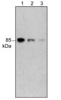
ARG20571 anti-PI3 Kinase p85 alpha antibody [M253] WB image
Western blot: A431 cells stained with ARG20571 anti-PI3 Kinase p85 alpha antibody [M253] at 1:1000 (Lane 1), 1:2000 (Lane 2), and 1:4000 (Lanes 3) dilutions.
-
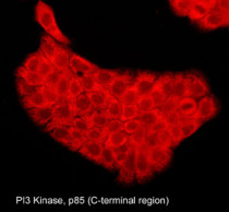
ARG20571 anti-PI3 Kinase p85 alpha antibody [M253] ICC/IF image
Immunocytochemistry: Aldehyde-fixed and NP-40-permeabilized A431 cells stained with ARG20571 anti-PI3 Kinase p85 alpha antibody [M253].


