ARG66996
anti-PERK antibody
anti-PERK antibody for ICC/IF,IHC-Formalin-fixed paraffin-embedded sections,Western blot and Human,Mouse
Overview
| Product Description | Rabbit Polyclonal antibody recognizes PERK |
|---|---|
| Tested Reactivity | Hu, Ms |
| Predict Reactivity | Rat |
| Tested Application | ICC/IF, IHC-P, WB |
| Host | Rabbit |
| Clonality | Polyclonal |
| Isotype | IgG |
| Target Name | PERK |
| Antigen Species | Human |
| Immunogen | Synthetic peptide corresponding to Human PERK. |
| Conjugation | Un-conjugated |
| Alternate Names | PRKR-like endoplasmic reticulum kinase; PERK; HsPEK; Eukaryotic translation initiation factor 2-alpha kinase 3; Pancreatic eIF2-alpha kinase; WRS; PEK; EC 2.7.11.1 |
Application Instructions
| Application Suggestion |
|
||||||||
|---|---|---|---|---|---|---|---|---|---|
| Application Note | * The dilutions indicate recommended starting dilutions and the optimal dilutions or concentrations should be determined by the scientist. | ||||||||
| Observed Size | ~ 130-140 kDa |
Properties
| Form | Liquid |
|---|---|
| Purification | Affinity purified. |
| Buffer | 100 mM Tris Glycine (pH 7.0), 0.025% ProClin 300 and 20% Glycerol. |
| Preservative | 0.025% ProClin 300 |
| Stabilizer | 20% Glycerol |
| Storage Instruction | For continuous use, store undiluted antibody at 2-8°C for up to a week. For long-term storage, aliquot and store at -20°C or below. Storage in frost free freezers is not recommended. Avoid repeated freeze/thaw cycles. Suggest spin the vial prior to opening. The antibody solution should be gently mixed before use. |
| Note | For laboratory research only, not for drug, diagnostic or other use. |
Bioinformation
| Database Links |
Swiss-port # Q9NZJ5 Human Eukaryotic translation initiation factor 2-alpha kinase 3 |
|---|---|
| Gene Symbol | EIF2AK3 |
| Gene Full Name | eukaryotic translation initiation factor 2-alpha kinase 3 |
| Background | The protein encoded by this gene phosphorylates the alpha subunit of eukaryotic translation-initiation factor 2, leading to its inactivation, and thus to a rapid reduction of translational initiation and repression of global protein synthesis. This protein is thought to modulate mitochondrial function. It is a type I membrane protein located in the endoplasmic reticulum (ER), where it is induced by ER stress caused by malfolded proteins. Mutations in this gene are associated with Wolcott-Rallison syndrome. [provided by RefSeq, Sep 2015] |
| Function | Phosphorylates the alpha subunit of eukaryotic translation-initiation factor 2 (EIF2), leading to its inactivation and thus to a rapid reduction of translational initiation and repression of global protein synthesis. Serves as a critical effector of unfolded protein response (UPR)-induced G1 growth arrest due to the loss of cyclin-D1 (CCND1). Involved in control of mitochondrial morphology and function (By similarity). [UniProt] |
| Calculated MW | 125 kDa |
| PTM | Oligomerization of the N-terminal ER luminal domain by ER stress promotes PERK trans-autophosphorylation of the C-terminal cytoplasmic kinase domain at multiple residues including Thr-982 on the kinase activation loop (By similarity). Autophosphorylated. Phosphorylated at Tyr-619 following endoplasmic reticulum stress, leading to activate its tyrosine-protein kinase activity. Dephosphorylated by PTPN1/TP1B, leading to inactivate its enzyme activity. N-glycosylated. ADP-ribosylated by PARP16 upon ER stress, which increases kinase activity. [UniProt] |
Images (5) Click the Picture to Zoom In
-
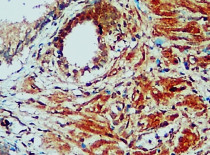
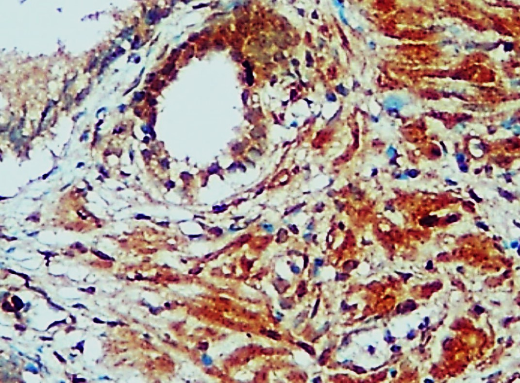
ARG66996 anti-PERK antibody IHC-p image
Immunohistochemistry: Formalin-fixed and paraffin-embedded human cancer tissue section tained with ARG66996 anti-PERK antibody.
-

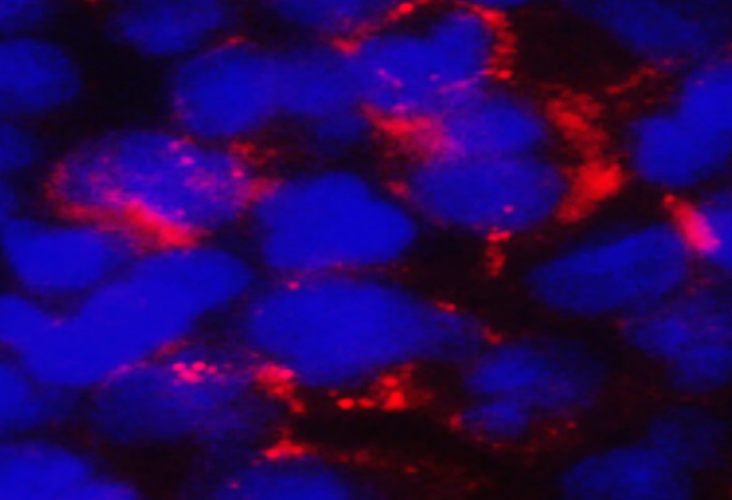
ARG66996 anti-PERK antibody ICC/IF image
Immunofluorescence: Formalin-fixed Raji cells were permeabilized with 0.1% NP-40 in TBS for 10 minutes and blocked with 5% BSA-PBS for 30 minutes at room temperature. Raji cell were stained with ARG66996 anti-PERK antibody.
-
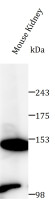
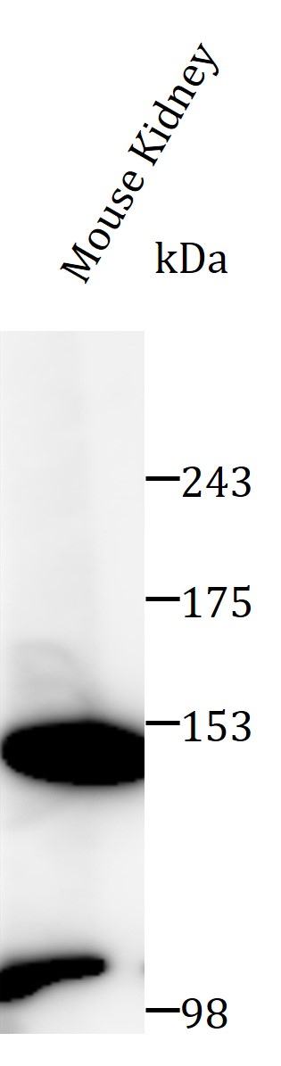
ARG66996 anti-PERK antibody WB image
Western blot: Mouse kidney stained with ARG66996 anti-PERK antibody at 1:500 dilution by 5% SDS-PAGE.
-
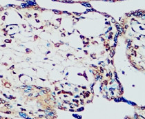
ARG66996 anti-PERK antibody IHC-p image
Immunohistochemistry: Formalin-fixed and paraffin-embedded human cancer tissue section tained with ARG66996 anti-PERK antibody.
-
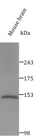
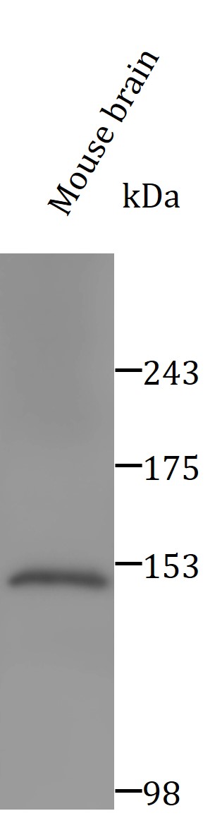
ARG66996 anti-PERK antibody WB image
Western blot: Mouse brain stained with ARG66996 anti-PERK antibody at 1:500 dilution by 5% SDS-PAGE.