ARG57836
anti-PEG10 antibody
anti-PEG10 antibody for IHC-Formalin-fixed paraffin-embedded sections,Western blot and Human,Mouse,Rat
Overview
| Product Description | Rabbit Polyclonal antibody recognizes PEG10 |
|---|---|
| Tested Reactivity | Hu, Ms, Rat |
| Tested Application | IHC-P, WB |
| Host | Rabbit |
| Clonality | Polyclonal |
| Isotype | IgG |
| Target Name | PEG10 |
| Antigen Species | Human |
| Immunogen | Recombinant protein of Human PEG10. |
| Conjugation | Un-conjugated |
| Alternate Names | MEF3L; Retrotransposon-derived gag-like polyprotein; RGAG3; Paternally expressed gene 10 protein; Myelin expression factor 3-like protein 1; MEF3-like protein 1; Mart2; Ty3/Gypsy-like protein; Retrotransposon-derived protein PEG10; Embryonal carcinoma differentiation-regulated protein; EDR; Mar2; Mammalian retrotransposon-derived protein 2; Retrotransposon gag domain-containing protein 3; HB-1 |
Application Instructions
| Application Suggestion |
|
||||||
|---|---|---|---|---|---|---|---|
| Application Note | * The dilutions indicate recommended starting dilutions and the optimal dilutions or concentrations should be determined by the scientist. | ||||||
| Positive Control | SW620 |
Properties
| Form | Liquid |
|---|---|
| Purification | Affinity purified. |
| Buffer | PBS (pH 7.3), 0.02% Sodium azide and 50% Glycerol. |
| Preservative | 0.02% Sodium azide |
| Stabilizer | 50% Glycerol |
| Storage Instruction | For continuous use, store undiluted antibody at 2-8°C for up to a week. For long-term storage, aliquot and store at -20°C. Storage in frost free freezers is not recommended. Avoid repeated freeze/thaw cycles. Suggest spin the vial prior to opening. The antibody solution should be gently mixed before use. |
| Note | For laboratory research only, not for drug, diagnostic or other use. |
Bioinformation
| Database Links |
Swiss-port # Q7TN75 Mouse Retrotransposon-derived protein PEG10 Swiss-port # Q86TG7 Human Retrotransposon-derived protein PEG10 |
|---|---|
| Gene Symbol | PEG10 |
| Gene Full Name | paternally expressed 10 |
| Background | This is a paternally expressed imprinted gene that is thought to have been derived from the Ty3/Gypsy family of retrotransposons. It contains two overlapping open reading frames, RF1 and RF2, and expresses two proteins: a shorter, gag-like protein (with a CCHC-type zinc finger domain) from RF1; and a longer, gag/pol-like fusion protein (with an additional aspartic protease motif) from RF1/RF2 by -1 translational frameshifting (-1 FS). While -1 FS has been observed in RNA viruses and transposons in both prokaryotes and eukaryotes, this gene represents the first example of -1 FS in a eukaryotic cellular gene. This gene is highly conserved across mammalian species and retains the heptanucleotide (GGGAAAC) and pseudoknot elements required for -1 FS. It is expressed in adult and embryonic tissues (most notably in placenta) and reported to have a role in cell proliferation, differentiation and apoptosis. Overexpression of this gene has been associated with several malignancies, such as hepatocellular carcinoma and B-cell lymphocytic leukemia. Knockout mice lacking this gene showed early embryonic lethality with placental defects, indicating the importance of this gene in embryonic development. Additional isoforms resulting from alternatively spliced transcript variants, and use of upstream non-AUG (CUG) start codon have been reported for this gene. [provided by RefSeq, Oct 2014] |
| Function | Prevents apoptosis in hepatocellular carcinoma (HCC) cells through interaction with SIAH1, a mediator of apoptosis. May also have a role in cell growth promotion and hepatoma formation. Inhibits the TGF-beta signaling by interacting with the TGF-beta receptor ALK1. When overexpressed, induces the formation of cellular extension, such as filipodia in association with ALK1. Involved at the immediate early stage of adipocyte differentiation (By similarity). May bind to the 5'-GCCTGTCTTT-3' DNA sequence of the MB1 domain in the myelin basic protein (MBP) promoter (By similarity). [UniProt] |
| Cellular Localization | Nucleus, Cytoplasm. [UniProt] |
| Calculated MW | 80 kDa |
| PTM | Isoform 1 undergoes proteolytic cleavage. [UniProt] |
Images (1) Click the Picture to Zoom In
-

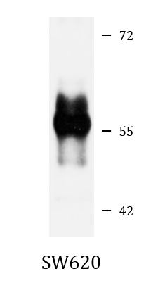
ARG57836 anti-PEG10 antibody WB image
Western blot: 25 µg of SW620 cell lysate stained with ARG57836 anti-PEG10 antibody at 1:1000 dilution.