ARG42967
anti-PEF1 / Peflin antibody
anti-PEF1 / Peflin antibody for IHC-Formalin-fixed paraffin-embedded sections,Western blot and Human,Mouse,Rat
Overview
| Product Description | Rabbit Polyclonal antibody recognizes PEF1 / Peflin |
|---|---|
| Tested Reactivity | Hu, Ms, Rat |
| Tested Application | IHC-P, WB |
| Host | Rabbit |
| Clonality | Polyclonal |
| Isotype | IgG |
| Target Name | PEF1 / Peflin |
| Antigen Species | Human |
| Immunogen | Recombinant protein corresponding to R170-L284 of Human PEF1 / Peflin. |
| Conjugation | Un-conjugated |
| Alternate Names | ABP32; PEF1A; Peflin; PEF protein with a long N-terminal hydrophobic domain; Penta-EF hand domain-containing protein 1 |
Application Instructions
| Application Suggestion |
|
||||||
|---|---|---|---|---|---|---|---|
| Application Note | IHC-P: Antigen Retrieval: Heat mediation was performed in Citrate buffer (pH 6.0) for 20 min. * The dilutions indicate recommended starting dilutions and the optimal dilutions or concentrations should be determined by the scientist. |
||||||
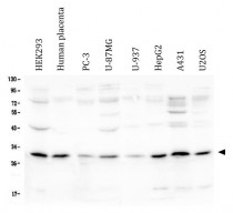
| Observed Size | ~ 30 kDa |
Properties
| Form | Liquid |
|---|---|
| Purification | Affinity purification with immunogen. |
| Buffer | 0.2% Na2HPO4, 0.9% NaCl, 0.05% Sodium azide and 4% Trehalose. |
| Preservative | 0.05% Sodium azide |
| Stabilizer | 4% Trehalose |
| Concentration | 0.5 mg/ml |
| Storage Instruction | For continuous use, store undiluted antibody at 2-8°C for up to a week. For long-term storage, aliquot and store at -20°C or below. Storage in frost free freezers is not recommended. Avoid repeated freeze/thaw cycles. Suggest spin the vial prior to opening. The antibody solution should be gently mixed before use. |
| Note | For laboratory research only, not for drug, diagnostic or other use. |
Bioinformation
| Database Links | |
|---|---|
| Gene Symbol | PEF1 |
| Gene Full Name | penta-EF-hand domain containing 1 |
| Background | This gene encodes a calcium-binding protein belonging to the penta-EF-hand protein family. The encoded protein has been shown to form a heterodimer with the programmed cell death 6 gene product and may modulate its function in Ca(2+) signaling. Alternative splicing results in multiple transcript variants and a pseudogene has been identified on chromosome 1. [provided by RefSeq, May 2010] |
| Function | Calcium-binding protein that acts as an adapter that bridges unrelated proteins or stabilizes weak protein-protein complexes in response to calcium. Together with PDCD6, acts as calcium-dependent adapter for the BCR(KLHL12) complex, a complex involved in endoplasmic reticulum (ER)-Golgi transport by regulating the size of COPII coats (PubMed:27716508). In response to cytosolic calcium increase, the heterodimer formed with PDCD6 interacts with, and bridges together the BCR(KLHL12) complex and SEC31 (SEC31A or SEC31B), promoting monoubiquitination of SEC31 and subsequent collagen export, which is required for neural crest specification (PubMed:27716508). Its role in the heterodimer formed with PDCD6 is however unclear: some evidence shows that PEF1 and PDCD6 work together and promote association between PDCD6 and SEC31 in presence of calcium (PubMed:27716508). Other reports show that PEF1 dissociates from PDCD6 in presence of calcium, and may act as a negative regulator of PDCD6 (PubMed:11278427). Also acts as a negative regulator of ER-Golgi transport; possibly by inhibiting interaction between PDCD6 and SEC31 (By similarity). [UniProt] |
| Cellular Localization | Cytoplasm. Endoplasmic reticulum. Membrane; Peripheral membrane protein. Cytoplasmic vesicle, COPII-coated vesicle membrane; Peripheral membrane protein. Note=Membrane-associated in the presence of Ca(2+) (PubMed:11278427). Localizes to endoplasmic reticulum exit site (ERES) (By similarity). [UniProt] |
| Calculated MW | 30 kDa |
| PTM | Ubiquitinated by the BCR(KLHL12) E3 ubiquitin ligase complex. [UniProt] |
Images (6) Click the Picture to Zoom In
-
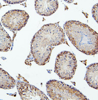
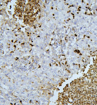
ARG42967 anti-PEF1 / Peflin antibody IHC-P image
Immunohistochemistry: Paraffin-embedded Mouse testis tissue. Antigen Retrieval: Heat mediation was performed in Citrate buffer (pH 6.0) for 20 min. The tissue section was blocked with 10% goat serum. The tissue section was then stained with ARG42967 anti-PEF1 / Peflin antibody at 1 µg/ml dilution, overnight at 4°C.
-
ARG42967 anti-PEF1 / Peflin antibody WB image
Western blot: 50 µg of sample under reducing conditions. HEK293, Human placenta, PC-3, U-87MG, U-937, HepG2, A431 and U2OS whole cell lysates stained with ARG42967 anti-PEF1 / Peflin antibody at 0.5 µg/ml dilution, overnight at 4°C.
-
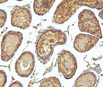
ARG42967 anti-PEF1 / Peflin antibody IHC-P image
Immunohistochemistry: Paraffin-embedded Rat testis tissue. Antigen Retrieval: Heat mediation was performed in Citrate buffer (pH 6.0) for 20 min. The tissue section was blocked with 10% goat serum. The tissue section was then stained with ARG42967 anti-PEF1 / Peflin antibody at 1 µg/ml dilution, overnight at 4°C.
-
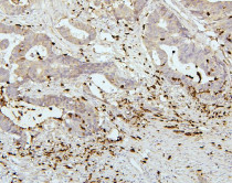
ARG42967 anti-PEF1 / Peflin antibody IHC-P image
Immunohistochemistry: Paraffin-embedded Human colon cancer tissue. Antigen Retrieval: Heat mediation was performed in Citrate buffer (pH 6.0) for 20 min. The tissue section was blocked with 10% goat serum. The tissue section was then stained with ARG42967 anti-PEF1 / Peflin antibody at 1 µg/ml dilution, overnight at 4°C.
-
ARG42967 anti-PEF1 / Peflin antibody IHC-P image
Immunohistochemistry: Paraffin-embedded Human lung cancer tissue. Antigen Retrieval: Heat mediation was performed in Citrate buffer (pH 6.0) for 20 min. The tissue section was blocked with 10% goat serum. The tissue section was then stained with ARG42967 anti-PEF1 / Peflin antibody at 1 µg/ml dilution, overnight at 4°C.
-
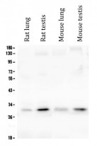
ARG42967 anti-PEF1 / Peflin antibody WB image
Western blot: 50 µg of sample under reducing conditions. Rat lung, Rat testis, Mouse lung and Mouse testis lysates stained with ARG42967 anti-PEF1 / Peflin antibody at 0.5 µg/ml dilution, overnight at 4°C.
