ARG66861
anti-PEA15 phospho (Ser104) antibody
anti-PEA15 phospho (Ser104) antibody for IHC-Formalin-fixed paraffin-embedded sections,Western blot and Human,Monkey
Overview
| Product Description | Rabbit Polyclonal antibody recognizes PEA15 phospho (Ser104) |
|---|---|
| Tested Reactivity | Hu, Mk |
| Predict Reactivity | Ms, Rat |
| Tested Application | IHC-P, WB |
| Specificity | This antibody detects endogenous levels of PEA15 protein only when phosphorylated at Ser104. |
| Host | Rabbit |
| Clonality | Polyclonal |
| Isotype | IgG |
| Target Name | PEA15 |
| Antigen Species | Human |
| Immunogen | Synthetic peptide around the phosphorylated Ser104 (aa. 70-119) of Human PEA15. |
| Conjugation | Un-conjugated |
| Alternate Names | MAT1H; MAT1; Astrocytic phosphoprotein PEA-15; HUMMAT1H; 15 kDa phosphoprotein enriched in astrocytes; PED; PEA-15; Phosphoprotein enriched in diabetes; HMAT1 |
Application Instructions
| Application Suggestion |
|
||||||
|---|---|---|---|---|---|---|---|
| Application Note | * The dilutions indicate recommended starting dilutions and the optimal dilutions or concentrations should be determined by the scientist. | ||||||
| Positive Control | COS7 | ||||||
| Observed Size | ~ 19 kDa |
Properties
| Form | Liquid |
|---|---|
| Purification | Affinity purification with immunogen. |
| Buffer | PBS, 0.02% Sodium azide, 50% Glycerol and 0.5% BSA. |
| Preservative | 0.02% Sodium azide |
| Stabilizer | 50% Glycerol and 0.5% BSA |
| Concentration | 1 mg/ml |
| Storage Instruction | For continuous use, store undiluted antibody at 2-8°C for up to a week. For long-term storage, aliquot and store at -20°C. Storage in frost free freezers is not recommended. Avoid repeated freeze/thaw cycles. Suggest spin the vial prior to opening. The antibody solution should be gently mixed before use. |
| Note | For laboratory research only, not for drug, diagnostic or other use. |
Bioinformation
| Database Links | |
|---|---|
| Gene Symbol | PEA15 |
| Gene Full Name | phosphoprotein enriched in astrocytes 15 |
| Background | This gene encodes a death effector domain-containing protein that functions as a negative regulator of apoptosis. The encoded protein is an endogenous substrate for protein kinase C. This protein is also overexpressed in type 2 diabetes mellitus, where it may contribute to insulin resistance in glucose uptake. Alternative splicing results in multiple transcript variants. [provided by RefSeq, Jul 2014] |
| Function | Blocks Ras-mediated inhibition of integrin activation and modulates the ERK MAP kinase cascade. Inhibits RPS6KA3 activities by retaining it in the cytoplasm (By similarity). Inhibits both TNFRSF6- and TNFRSF1A-mediated CASP8 activity and apoptosis. Regulates glucose transport by controlling both the content of SLC2A1 glucose transporters on the plasma membrane and the insulin-dependent trafficking of SLC2A4 from the cell interior to the surface. [UniProt] |
| Cellular Localization | Cytoplasm. Note=Associated with microtubules. [UniProt] |
| Calculated MW | 15 kDa |
| PTM | Phosphorylated by protein kinase C and calcium-calmodulin-dependent protein kinase. These phosphorylation events are modulated by neurotransmitters or hormones. [UniProt] |
Images (2) Click the Picture to Zoom In
-
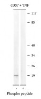
ARG66861 anti-PEA15 phospho (Ser104) antibody WB image
Western blot: COS7 cells treated with TNF at 20 ng/ml for 5 min. Cell lysates were stained with ARG66861 anti-PEA15 phospho (Ser104) antibody. The lane on the right is blocked with the phospho peptide.
-
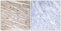
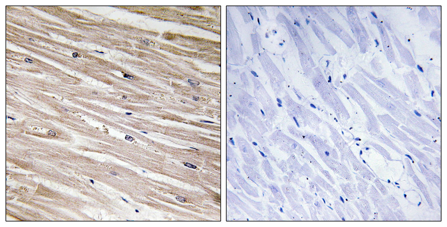
ARG66861 anti-PEA15 phospho (Ser104) antibody IHC-P image
Immunohistochemistry: Paraffin-embedded Human heart tissue stained with ARG66861 anti-PEA15 phospho (Ser104) antibody. The picture on the right is blocked with the phospho peptide.