ARG43792
anti-PDX1 antibody
anti-PDX1 antibody for Flow cytometry,Western blot and Human
Overview
| Product Description | Rabbit Polyclonal antibody recognizes PDX1 |
|---|---|
| Tested Reactivity | Hu |
| Tested Application | FACS, WB |
| Host | Rabbit |
| Clonality | Polyclonal |
| Isotype | IgG |
| Target Name | PDX1 |
| Antigen Species | Human |
| Immunogen | Recombinant protein corresponding to Human PDX1. |
| Conjugation | Un-conjugated |
| Protein Full Name | Pancreas/duodenum homeobox protein 1 |
| Alternate Names | IPF1; Glucose-sensitive factor; Somatostatin-transactivating factor 1; Pancreas/duodenum homeobox protein 1; GSF; Insulin upstream factor 1; IUF-1; IUF1; IDX-1; Insulin promoter factor 1; MODY4; PDX-1; PAGEN1; IPF-1; STF-1; Islet/duodenum homeobox-1 |
Application Instructions
| Application Suggestion |
|
||||||
|---|---|---|---|---|---|---|---|
| Application Note | * The dilutions indicate recommended starting dilutions and the optimal dilutions or concentrations should be determined by the scientist. | ||||||
| Positive Control | Mouse pancreas | ||||||
| Observed Size | ~ 40 kDa |
Properties
| Form | Liquid |
|---|---|
| Purification | Affinity purification with immunogen. |
| Buffer | 0.2% Na2HPO4, 0.9% NaCl, 0.01% Sodium azide and 4% Trehalose. |
| Preservative | 0.01% Sodium azide |
| Stabilizer | 4% Trehalose |
| Concentration | 0.5 mg/ml |
| Storage Instruction | For continuous use, store undiluted antibody at 2-8°C for up to a week. For long-term storage, aliquot and store at -20°C or below. Storage in frost free freezers is not recommended. Avoid repeated freeze/thaw cycles. Suggest spin the vial prior to opening. The antibody solution should be gently mixed before use. |
| Note | For laboratory research only, not for drug, diagnostic or other use. |
Bioinformation
| Database Links |
Swiss-port # P52945 Human Pancreas/duodenum homeobox protein 1 |
|---|---|
| Gene Symbol | PDX1 |
| Gene Full Name | pancreatic and duodenal homeobox 1 |
| Background | The protein encoded by this gene is a transcriptional activator of several genes, including insulin, somatostatin, glucokinase, islet amyloid polypeptide, and glucose transporter type 2. The encoded nuclear protein is involved in the early development of the pancreas and plays a major role in glucose-dependent regulation of insulin gene expression. Defects in this gene are a cause of pancreatic agenesis, which can lead to early-onset insulin-dependent diabetes mellitus (NIDDM), as well as maturity onset diabetes of the young type 4 (MODY4). [provided by RefSeq, Jul 2008] |
| Function | Activates insulin, somatostatin, glucokinase, islet amyloid polypeptide and glucose transporter type 2 gene transcription. Particularly involved in glucose-dependent regulation of insulin gene transcription. As part of a PDX1:PBX1b:MEIS2b complex in pancreatic acinar cells is involved in the transcriptional activation of the ELA1 enhancer; the complex binds to the enhancer B element and cooperates with the transcription factor 1 complex (PTF1) bound to the enhancer A element. Binds preferentially the DNA motif 5'-[CT]TAAT[TG]-3'. During development, specifies the early pancreatic epithelium, permitting its proliferation, branching and subsequent differentiation. At adult stage, required for maintaining the hormone-producing phenotype of the beta-cell. [UniProt] |
| Cellular Localization | Nucleus. Cytoplasm, cytosol. [UniProt] |
| Calculated MW | 31 kDa |
| PTM | Phosphorylated by the SAPK2 pathway at high intracellular glucose concentration. Phosphorylated by HIPK2 on Ser-268 upon glucose accumulation. This phosphorylation mediates subnuclear localization shifting. Phosphorylation by PASK may lead to translocation into the cytosol (By similarity). [UniProt] |
Images (2) Click the Picture to Zoom In
-
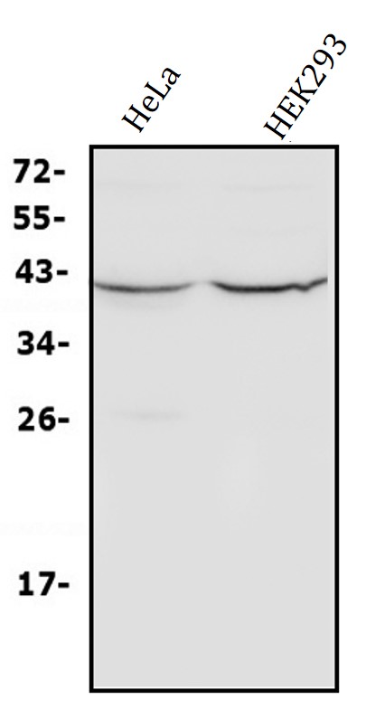
ARG43792 anti-PDX1 antibody WB image
Western blot: HeLa and HEK293 cell lysates stained with ARG43792 anti-PDX1 antibody.
-
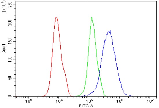
ARG43792 anti-PDX1 antibody FACS image
Flow Cytometry: 293T cells were blocked with 10% normal goat serum and then stained with ARG43792 anti-PDX1 antibody (blue) at 1 µg/10^6 cells for 30 min at 20°C, followed by incubation with DyLight®488 labelled secondary antibody. Isotype control antibody (green) was Rabbit IgG (1 µg/10^6 cells) used under the same conditions. Unlabelled sample (red) was also used as a control.