ARG40409
anti-PDP1 antibody
anti-PDP1 antibody for IHC-Formalin-fixed paraffin-embedded sections,Western blot and Human
Overview
| Product Description | Rabbit Polyclonal antibody recognizes PDP1 |
|---|---|
| Tested Reactivity | Hu |
| Tested Application | IHC-P, WB |
| Host | Rabbit |
| Clonality | Polyclonal |
| Isotype | IgG |
| Target Name | PDP1 |
| Antigen Species | Human |
| Immunogen | Fusion protein of Human PDP1. |
| Conjugation | Un-conjugated |
| Alternate Names | PDPC; PDPC 1; PDP; EC 3.1.3.43; Protein phosphatase 2C; PDH; PDP 1; PPM2C; [Pyruvate dehydrogenase [acetyl-transferring]]-phosphatase 1, mitochondrial; Pyruvate dehydrogenase phosphatase catalytic subunit 1 |
Application Instructions
| Application Suggestion |
|
||||||
|---|---|---|---|---|---|---|---|
| Application Note | * The dilutions indicate recommended starting dilutions and the optimal dilutions or concentrations should be determined by the scientist. | ||||||
| Positive Control | WB: Human bladder carcinoma tissue. IHC-P: Human thyroid cancer and Human cervical cancer. |
||||||
| Observed Size | ~ 60 kDa |
Properties
| Form | Liquid |
|---|---|
| Purification | Affinity purification with immunogen. |
| Buffer | PBS (pH 7.4), 0.05% Sodium azide and 40% Glycerol. |
| Preservative | 0.05% Sodium azide |
| Stabilizer | 40% Glycerol |
| Concentration | 1.3 mg/ml |
| Storage Instruction | For continuous use, store undiluted antibody at 2-8°C for up to a week. For long-term storage, aliquot and store at -20°C. Storage in frost free freezers is not recommended. Avoid repeated freeze/thaw cycles. Suggest spin the vial prior to opening. The antibody solution should be gently mixed before use. |
| Note | For laboratory research only, not for drug, diagnostic or other use. |
Bioinformation
| Database Links |
Swiss-port # Q9P0J1 Human [Pyruvate dehydrogenase [acetyl-transferring]]-phosphatase 1, mitochondria |
|---|---|
| Gene Symbol | PDP1 |
| Gene Full Name | pyruvate dehyrogenase phosphatase catalytic subunit 1 |
| Background | Pyruvate dehydrogenase (E1) is one of the three components (E1, E2, and E3) of the large pyruvate dehydrogenase complex. Pyruvate dehydrogenase kinases catalyze phosphorylation of serine residues of E1 to inactivate the E1 component and inhibit the complex. Pyruvate dehydrogenase phosphatases catalyze the dephosphorylation and activation of the E1 component to reverse the effects of pyruvate dehydrogenase kinases. Pyruvate dehydrogenase phosphatase is a heterodimer consisting of catalytic and regulatory subunits. Two catalytic subunits have been reported; one is predominantly expressed in skeletal muscle and another one is is much more abundant in the liver. The catalytic subunit, encoded by this gene, is the former, and belongs to the protein phosphatase 2C (PP2C) superfamily. Along with the pyruvate dehydrogenase complex and pyruvate dehydrogenase kinases, this enzyme is located in the mitochondrial matrix. Mutation in this gene causes pyruvate dehydrogenase phosphatase deficiency. Multiple alternatively spliced transcript variants encoding different isoforms have been identified.[provided by RefSeq, Jun 2009] |
| Function | Catalyzes the dephosphorylation and concomitant reactivation of the alpha subunit of the E1 component of the pyruvate dehydrogenase complex. [UniProt] |
| Cellular Localization | Mitochondrion matrix. [UniProt] |
| Calculated MW | 61 kDa |
Images (3) Click the Picture to Zoom In
-
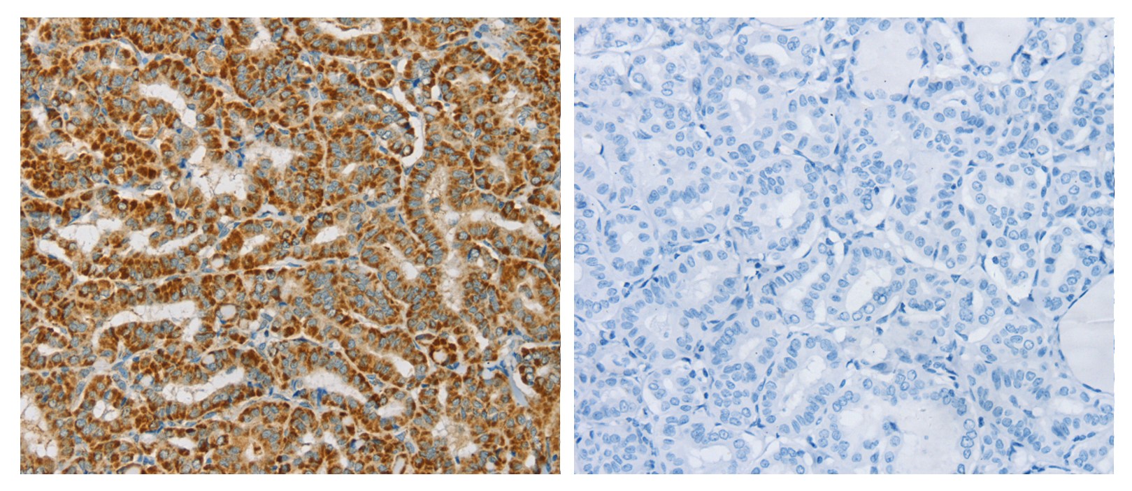
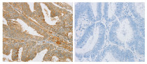
ARG40409 anti-PDP1 antibody IHC-P image
Immunohistochemistry: Paraffin-embedded Human thyroid cancer tissue stained with ARG40409 anti-PDP1 antibody (left) at 1:25 dilution, or the same antibody pre-incubated with fusion protein (right).
-
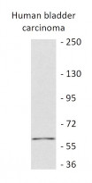
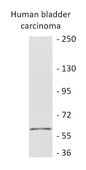
ARG40409 anti-PDP1 antibody WB image
Western blot: 40 µg of Human bladder carcinoma tissue lysate stained with ARG40409 anti-PDP1 antibody at 1:300 dilution.
-
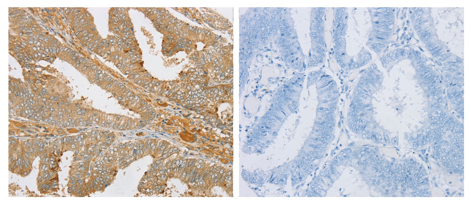
ARG40409 anti-PDP1 antibody IHC-P image
Immunohistochemistry: Paraffin-embedded Human cervical cancer tissue stained with ARG40409 anti-PDP1 antibody (left) at 1:25 dilution, or the same antibody pre-incubated with fusion protein (right).