ARG45519
anti-PDK3 antibody
anti-PDK3 antibody for Flow cytometry,ICC/IF,IHC-Formalin-fixed paraffin-embedded sections,Western blot and Human,Mouse,Rat
Overview
| Product Description | Rabbit Polyclonal antibody recognizes PDK3 |
|---|---|
| Tested Reactivity | Hu, Ms, Rat |
| Tested Application | FACS, ICC/IF, IHC-P, WB |
| Host | Rabbit |
| Clonality | Polyclonal |
| Isotype | IgG |
| Target Name | PDK3 |
| Antigen Species | Human |
| Immunogen | Recombinant protein containing to human PDK3. |
| Conjugation | Un-conjugated |
| Alternate Names | PDK3; pyruvate dehydrogenase kinase, isozyme 3; CMTX6; GS1-358P8.4; [Pyruvate dehydrogenase (acetyl-transferring)] kinase isozyme 3, mitochondrial; EC 2.7.11.2; Pyruvate dehydrogenase kinase isoform 3 |
Application Instructions
| Application Suggestion |
|
||||||||||
|---|---|---|---|---|---|---|---|---|---|---|---|
| Application Note | * The dilutions indicate recommended starting dilutions and the optimal dilutions or concentrations should be determined by the scientist. | ||||||||||
| Observed Size | 47 kDa |
Properties
| Form | Liquid |
|---|---|
| Purification | Affinity purified |
| Buffer | 0.2% Na2HPO4, 0.9% NaCl and 4% Trehalose. |
| Stabilizer | 4% Trehalose |
| Concentration | 0.5 mg/ml |
| Storage Instruction | For continuous use, store undiluted antibody at 2-8°C for up to a week. For long-term storage, aliquot and store at -20°C or below. Storage in frost free freezers is not recommended. Avoid repeated freeze/thaw cycles. Suggest spin the vial prior to opening. The antibody solution should be gently mixed before use. |
| Note | For laboratory research only, not for drug, diagnostic or other use. |
Bioinformation
| Database Links |
Swiss-port # Q15120 Human [Pyruvate dehydrogenase (acetyl-transferring)] kinase isozyme 3, mitochond Swiss-port # Q922H2 Mouse [Pyruvate dehydrogenase (acetyl-transferring)] kinase isozyme 3, mitochond |
|---|---|
| Gene Symbol | PDK3 |
| Gene Full Name | pyruvate dehydrogenase kinase, isozyme 3 |
| Background | The pyruvate dehydrogenase (PDH) complex is a nuclear-encoded mitochondrial multienzyme complex that catalyzes the overall conversion of pyruvate to acetyl-CoA and CO(2). It provides the primary link between glycolysis and the tricarboxylic acid (TCA) cycle, and thus is one of the major enzymes responsible for the regulation of glucose metabolism. The enzymatic activity of PDH is regulated by a phosphorylation/dephosphorylation cycle, and phosphorylation results in inactivation of PDH. The protein encoded by this gene is one of the three pyruvate dehydrogenase kinases that inhibits the PDH complex by phosphorylation of the E1 alpha subunit. This gene is predominantly expressed in the heart and skeletal muscles. Alternatively spliced transcript variants encoding different isoforms have been found for this gene. [provided by RefSeq, Mar 2010] |
| Function | Inhibits pyruvate dehydrogenase activity by phosphorylation of the E1 subunit PDHA1, and thereby regulates glucose metabolism and aerobic respiration. Can also phosphorylate PDHA2. Decreases glucose utilization and increases fat metabolism in response to prolonged fasting, and as adaptation to a high-fat diet. Plays a role in glucose homeostasis and in maintaining normal blood glucose levels in function of nutrient levels and under starvation. Plays a role in the generation of reactive oxygen species. [UniProt] |
| Cellular Localization | Mitochondrion matrix. [UniProt] |
| Calculated MW | 47 kDa |
Images (8) Click the Picture to Zoom In
-
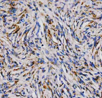
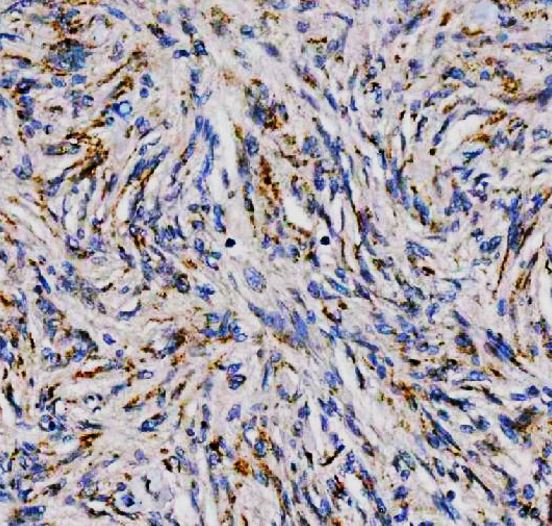
ARG45519 anti-PDK3 antibody IHC-P image
Immunohistochemistry: Human meningioma stained with ARG45519 anti-PDK3 antibody at 2 μg/ml dilution.
-
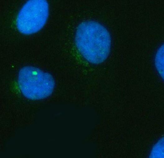
ARG45519 anti-PDK3 antibody ICC/IF image
Immunofluorescence: Hela stained with ARG45519 anti-PDK3 antibody at 5 μg/ml dilution.
-
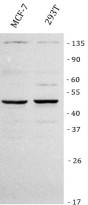
ARG45519 anti-PDK3 antibody WB image
Western blot: MCF-7 and 293T stained with ARG45519 anti-PDK3 antibody at 0.5 μg/ml dilution.
-

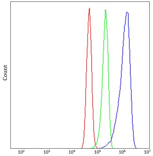
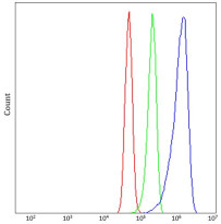
ARG45519 anti-PDK3 antibody FACS image
Flow Cytometry: U251 stained with ARG45519 anti-PDK3 antibody at 1 µg/10^6 cells dilution.
-
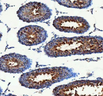
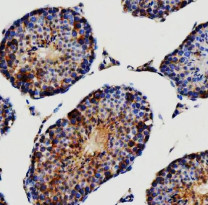
ARG45519 anti-PDK3 antibody IHC-P image
Immunohistochemistry: Rat testis stained with ARG45519 anti-PDK3 antibody at 2 μg/ml dilution.
-
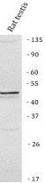
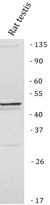
ARG45519 anti-PDK3 antibody WB image
Western blot: Rat testis stained with ARG45519 anti-PDK3 antibody at 0.5 μg/ml dilution.
-
ARG45519 anti-PDK3 antibody IHC-P image
Immunohistochemistry: Mouse testis stained with ARG45519 anti-PDK3 antibody at 2 μg/ml dilution.
-
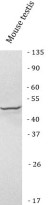
ARG45519 anti-PDK3 antibody WB image
Western blot: Mouse testis stained with ARG45519 anti-PDK3 antibody at 0.5 μg/ml dilution.