ARG55843
anti-PDIA6 antibody
anti-PDIA6 antibody for Flow cytometry,ICC/IF,IHC-Formalin-fixed paraffin-embedded sections,Western blot and Human,Mouse,Rat
Overview
| Product Description | Rabbit Polyclonal antibody recognizes PDIA6 |
|---|---|
| Tested Reactivity | Hu, Ms, Rat |
| Tested Application | FACS, ICC/IF, IHC-P, WB |
| Host | Rabbit |
| Clonality | Polyclonal |
| Isotype | IgG |
| Target Name | PDIA6 |
| Antigen Species | Human |
| Immunogen | KLH-conjugated synthetic peptide corresponding to aa. 144-172 (Center) of Human PDIA6. |
| Conjugation | Un-conjugated |
| Alternate Names | Protein disulfide isomerase P5; ERP5; Thioredoxin domain-containing protein 7; P5; ER protein 5; Endoplasmic reticulum protein 5; ERp5; EC 5.3.4.1; Protein disulfide-isomerase A6; TXNDC7 |
Application Instructions
| Application Suggestion |
|
||||||||||
|---|---|---|---|---|---|---|---|---|---|---|---|
| Application Note | * The dilutions indicate recommended starting dilutions and the optimal dilutions or concentrations should be determined by the scientist. | ||||||||||
| Positive Control | K562 |
Properties
| Form | Liquid |
|---|---|
| Purification | This antibody is prepared by Saturated Ammonium Sulfate (SAS) precipitation followed by dialysis against PBS. |
| Buffer | PBS and 0.09% (W/V) Sodium azide |
| Preservative | 0.09% (W/V) Sodium azide |
| Storage Instruction | For continuous use, store undiluted antibody at 2-8°C for up to a week. For long-term storage, aliquot and store at -20°C or below. Storage in frost free freezers is not recommended. Avoid repeated freeze/thaw cycles. Suggest spin the vial prior to opening. The antibody solution should be gently mixed before use. |
| Note | For laboratory research only, not for drug, diagnostic or other use. |
Bioinformation
| Database Links | |
|---|---|
| Gene Symbol | PDIA6 |
| Gene Full Name | protein disulfide isomerase family A, member 6 |
| Background | Protein disulfide isomerases (EC 5.3.4.1), such as PDIA6, are endoplasmic reticulum (ER) resident proteins that catalyze formation, reduction, and isomerization of disulfide bonds in proteins and are thought to play a role in folding of disulfide-bonded proteins (Hayano and Kikuchi, 1995 [PubMed 7590364]).[supplied by OMIM, Mar 2008] |
| Function | May function as a chaperone that inhibits aggregation of misfolded proteins. Plays a role in platelet aggregation and activation by agonists such as convulxin, collagen and thrombin. [UniProt] |
| Cellular Localization | Endoplasmic reticulum lumen {ECO:0000255|PROSITE-ProRule:PRU10138}. Cell membrane. Melanosome Note=Identified by mass spectrometry in melanosome fractions from stage I to stage IV |
| Calculated MW | 48 kDa |
Images (4) Click the Picture to Zoom In
-
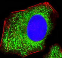
ARG55843 anti-PDIA6 antibody ICC/IF image
Immunofluorescence: HepG2 cells stained with ARG55843 anti-PDIA6 antibody (green) at 1:100 dilution. Cytoplasmic actin was counterstained with Alexa Fluor® 555 conjugated with Phalloidin (red). DAPI (blue) for nuclear staining.
-
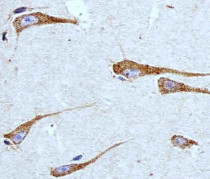
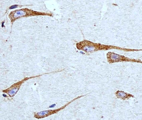
ARG55843 anti-PDIA6 antibody IHC-P image
Immunohistochemistry: Paraffin-embedded Human brain tissue stained with ARG55843 anti-PDIA6 antibody at 1:100 dilution.
-
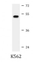
ARG55843 anti-PDIA6 antibody WB image
Western blot: 35 µg of K562 cell lysate stained with ARG55843 anti-PDIA6 antibody at 1:1000 dilution.
-
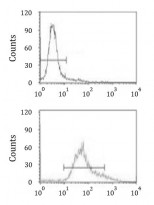
ARG55843 anti-PDIA6 antibody FACS image
Flow Cytometry: HeLa cells stained with ARG55843 anti-PDIA6 antibody (bottom histogram) or without primary antibody control (top histogram), followed by incubation with FITC labelled secondary antibody.