ARG43461
anti-PDHX antibody
anti-PDHX antibody for Immunoprecipitation,Western blot and Human,Mouse,Rat
Overview
| Product Description | Rabbit Polyclonal antibody recognizes PDHX. |
|---|---|
| Tested Reactivity | Hu, Ms, Rat |
| Tested Application | IP, WB |
| Host | Rabbit |
| Clonality | Polyclonal |
| Isotype | IgG |
| Target Name | PDHX |
| Antigen Species | Human |
| Immunogen | Purified recombinant protein corresponding to human PDHX. |
| Conjugation | Un-conjugated |
| Protein Full Name | Pyruvate dehydrogenase protein X component, mitochondrial |
| Alternate Names | E3BP; OPDX; PDX1; proX; DLDBP |
Application Instructions
| Application Suggestion |
|
||||||
|---|---|---|---|---|---|---|---|
| Application Note | * The dilutions indicate recommended starting dilutions and the optimal dilutions or concentrations should be determined by the scientist. |
Properties
| Form | Liquid |
|---|---|
| Purification | Affinity purified. |
| Buffer | PBS (pH 7.3), 0.02% Sodium azide and 50% Glycerol. |
| Preservative | 0.02% Sodium azide |
| Stabilizer | 50% Glycerol |
| Storage Instruction | For continuous use, store undiluted antibody at 2-8°C for up to a week. For long-term storage, aliquot and store at -20°C or below. Storage in frost free freezers is not recommended. Avoid repeated freeze/thaw cycles. Suggest spin the vial prior to opening. The antibody solution should be gently mixed before use. |
| Note | For laboratory research only, not for drug, diagnostic or other use. |
Bioinformation
| Database Links |
Swiss-port # O00330 Human Pyruvate dehydrogenase protein X component, mitochondrial Swiss-port # Q8BKZ9 Mouse Pyruvate dehydrogenase protein X component, mitochondrial |
|---|---|
| Gene Symbol | PDHX |
| Gene Full Name | pyruvate dehydrogenase complex, component X |
| Background | The pyruvate dehydrogenase (PDH) complex is located in the mitochondrial matrix and catalyzes the conversion of pyruvate to acetyl coenzyme A. The PDH complex thereby links glycolysis to Krebs cycle. The PDH complex contains three catalytic subunits, E1, E2, and E3, two regulatory subunits, E1 kinase and E1 phosphatase, and a non-catalytic subunit, E3 binding protein (E3BP). This gene encodes the E3 binding protein subunit; also known as component X of the pyruvate dehydrogenase complex. This protein tethers E3 dimers to the E2 core of the PDH complex. Defects in this gene are a cause of pyruvate dehydrogenase deficiency which results in neurological dysfunction and lactic acidosis in infancy and early childhood. This protein is also a minor antigen for antimitochondrial antibodies. These autoantibodies are present in nearly 95% of patients with the autoimmune liver disease primary biliary cirrhosis (PBC). In PBC, activated T lymphocytes attack and destroy epithelial cells in the bile duct where this protein is abnormally distributed and overexpressed. PBC eventually leads to cirrhosis and liver failure. Alternative splicing results in multiple transcript variants encoding distinct isoforms.[provided by RefSeq, Oct 2009] |
| Function | Required for anchoring dihydrolipoamide dehydrogenase (E3) to the dihydrolipoamide transacetylase (E2) core of the pyruvate dehydrogenase complexes of eukaryotes. This specific binding is essential for a functional PDH complex. [UniProt] |
Images (3) Click the Picture to Zoom In
-
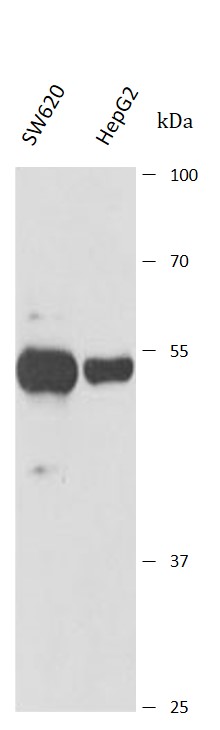
ARG43461 anti-PDHX antibody WB image
Western blot: SW620 and HepG2 stained with ARG43461 anti-PDHX antibody at 1:1000 dilution.
-
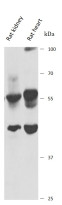
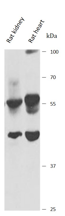
ARG43461 anti-PDHX antibody WB image
Western blot: Rat kidney and Rat heart stained with ARG43461 anti-PDHX antibody at 1:1000 dilution.
-
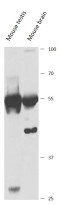
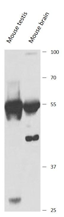
ARG43461 anti-PDHX antibody WB image
Western blot: Mouse testis and Mouse brain stained with ARG43461 anti-PDHX antibody at 1:1000 dilution.