ARG63878
anti-PDE4D antibody
anti-PDE4D antibody for ELISA,IHC-Formalin-fixed paraffin-embedded sections,Western blot and Human
Signaling Transduction antibody
Overview
| Product Description | Goat Polyclonal antibody recognizes PDE4D |
|---|---|
| Tested Reactivity | Hu |
| Tested Application | ELISA, IHC-P, WB |
| Specificity | This antiobody is expected to recognize all reported isoforms. |
| Host | Goat |
| Clonality | Polyclonal |
| Isotype | IgG |
| Target Name | PDE4D |
| Antigen Species | Human |
| Immunogen | QPEACVIDDRSPDT |
| Conjugation | Un-conjugated |
| Alternate Names | EC 3.1.4.53; STRK1; DPDE3; PDE43; ACRDYS2; PDE4DN2; cAMP-specific 3',5'-cyclic phosphodiesterase 4D; HSPDE4D |
Application Instructions
| Application Suggestion |
|
||||||||
|---|---|---|---|---|---|---|---|---|---|
| Application Note | WB: Recommend incubate at RT for 1h. IHC-P: Antigen Retrieval: Steam tissue section in Citrate buffer (pH 6.0). * The dilutions indicate recommended starting dilutions and the optimal dilutions or concentrations should be determined by the scientist. |
Properties
| Form | Liquid |
|---|---|
| Purification | Purified from goat serum by antigen affinity chromatography. |
| Buffer | Tris saline (pH 7.3), 0.02% Sodium azide and 0.5% BSA. |
| Preservative | 0.02% Sodium azide |
| Stabilizer | 0.5% BSA |
| Concentration | 0.5 mg/ml |
| Storage Instruction | For continuous use, store undiluted antibody at 2-8°C for up to a week. For long-term storage, aliquot and store at -20°C or below. Storage in frost free freezers is not recommended. Avoid repeated freeze/thaw cycles. Suggest spin the vial prior to opening. The antibody solution should be gently mixed before use. |
| Note | For laboratory research only, not for drug, diagnostic or other use. |
Bioinformation
| Database Links |
Swiss-port # Q08499 Human cAMP-specific 3',5'-cyclic phosphodiesterase 4D |
|---|---|
| Background | This gene encodes one of four mammalian counterparts to the fruit fly 'dunce' gene. The encoded protein has 3',5'-cyclic-AMP phosphodiesterase activity and degrades cAMP, which acts as a signal transduction molecule in multiple cell types. This gene uses different promoters to generate multiple alternatively spliced transcript variants that encode functional proteins.[provided by RefSeq, Sep 2009] |
| Research Area | Signaling Transduction antibody |
| Calculated MW | 91 kDa |
| PTM | Long isoforms that share a conserved PKA phosphorylation site in the N-terminus are activated by PKA through phosphorylation (By similarity). Isoform 3 and isoform 7 are activated by phosphorylation (in vitro), but not isoform 6. Isoform N3 and isoform 12 are phosphorylated on Ser-49, Ser-51, Ser-55 and Ser-59. Sumoylation of long isoforms by PIAS4 augments their activation by PKA phosphorylation and represses their inhibition by ERK phosphorylation. |
Images (4) Click the Picture to Zoom In
-
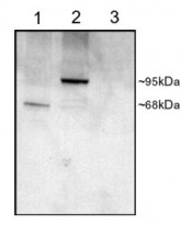
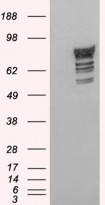
ARG63878 anti-PDE4D antibody WB image
Western Blot: COS cell lysates (25µg protein): transfected with Human PDE4D1 (1), transfected with Human PDE4D3 (2), untransfected (3) stained with ARG63878 anti-PDE4D antibody at 1 µg/ml dilution.
-
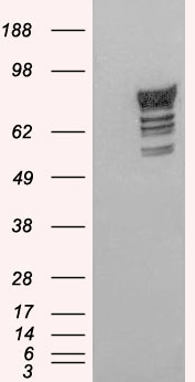
ARG63878 anti-PDE4D antibody WB image
Western Blot: 1). Mock transfection; 2) Human PDE4D2 (RC212410) expressing plasmid transfected HEK293 cell lysate standed with ARG63878 anti-PDE4D antibody
-
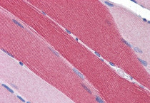
ARG63878 anti-PDE4D antibody IHC-P image
Immunohistochemistry: Paraffin-embedded Human skeletal muscle tissue. Antigen Retrieval: Steam tissue section in Citrate buffer (pH 6.0). The tissue section was stained with ARG63878 anti-PDE4D antibody at 5 µg/ml dilution followed by AP-staining.
-
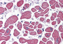
ARG63878 anti-PDE4D antibody IHC-P image
Immunohistochemistry: Paraffin-embedded Human heart tissue. Antigen Retrieval: Steam tissue section in Citrate buffer (pH 6.0). The tissue section was stained with ARG63878 anti-PDE4D antibody at 5 µg/ml dilution followed by AP-staining.