ARG55447
anti-PCSK9 antibody
anti-PCSK9 antibody for Flow cytometry,IHC-Formalin-fixed paraffin-embedded sections,Western blot and Human
Cell Biology and Cellular Response antibody; Developmental Biology antibody; Metabolism antibody; Signaling Transduction antibody
Overview
| Product Description | Rabbit Polyclonal antibody recognizes PCSK9 |
|---|---|
| Tested Reactivity | Hu |
| Tested Application | FACS, IHC-P, WB |
| Host | Rabbit |
| Clonality | Polyclonal |
| Isotype | IgG |
| Target Name | PCSK9 |
| Antigen Species | Human |
| Immunogen | KLH-conjugated synthetic peptide corresponding to aa. 144-173 (N-terminus) of Human PCSK9. |
| Conjugation | Un-conjugated |
| Alternate Names | PC9; Subtilisin/kexin-like protease PC9; Proprotein convertase 9; Proprotein convertase subtilisin/kexin type 9; Neural apoptosis-regulated convertase 1; FH3; EC 3.4.21.-; HCHOLA3; NARC1; LDLCQ1; NARC-1 |
Application Instructions
| Application Suggestion |
|
||||||||
|---|---|---|---|---|---|---|---|---|---|
| Application Note | * The dilutions indicate recommended starting dilutions and the optimal dilutions or concentrations should be determined by the scientist. | ||||||||
| Positive Control | HepG2 |
Properties
| Form | Liquid |
|---|---|
| Purification | Purification with Protein A and immunogen peptide. |
| Buffer | PBS and 0.09% (W/V) Sodium azide |
| Preservative | 0.09% (W/V) Sodium azide |
| Storage Instruction | For continuous use, store undiluted antibody at 2-8°C for up to a week. For long-term storage, aliquot and store at -20°C or below. Storage in frost free freezers is not recommended. Avoid repeated freeze/thaw cycles. Suggest spin the vial prior to opening. The antibody solution should be gently mixed before use. |
| Note | For laboratory research only, not for drug, diagnostic or other use. |
Bioinformation
| Database Links |
Swiss-port # Q8NBP7 Human Proprotein convertase subtilisin/kexin type 9 |
|---|---|
| Gene Symbol | PCSK9 |
| Gene Full Name | proprotein convertase subtilisin/kexin type 9 |
| Background | This gene encodes a member of the subtilisin-like proprotein convertase family, which includes proteases that process protein and peptide precursors trafficking through regulated or constitutive branches of the secretory pathway. The encoded protein undergoes an autocatalytic processing event with its prosegment in the ER and is constitutively secreted as an inactive protease into the extracellular matrix and trans-Golgi network. It is expressed in liver, intestine and kidney tissues and escorts specific receptors for lysosomal degradation. It plays a role in cholesterol and fatty acid metabolism. Mutations in this gene have been associated with autosomal dominant familial hypercholesterolemia. Alternative splicing results in multiple transcript variants. [provided by RefSeq, Feb 2014] |
| Function | Crucial player in the regulation of plasma cholesterol homeostasis. Binds to low-density lipid receptor family members: low density lipoprotein receptor (LDLR), very low density lipoprotein receptor (VLDLR), apolipoprotein E receptor (LRP1/APOER) and apolipoprotein receptor 2 (LRP8/APOER2), and promotes their degradation in intracellular acidic compartments. Acts via a non-proteolytic mechanism to enhance the degradation of the hepatic LDLR through a clathrin LDLRAP1/ARH-mediated pathway. May prevent the recycling of LDLR from endosomes to the cell surface or direct it to lysosomes for degradation. Can induce ubiquitination of LDLR leading to its subsequent degradation. Inhibits intracellular degradation of APOB via the autophagosome/lysosome pathway in a LDLR-independent manner. Involved in the disposal of non-acetylated intermediates of BACE1 in the early secretory pathway. Inhibits epithelial Na(+) channel (ENaC)-mediated Na(+) absorption by reducing ENaC surface expression primarily by increasing its proteasomal degradation. Regulates neuronal apoptosis via modulation of LRP8/APOER2 levels and related anti-apoptotic signaling pathways. [UniProt] |
| Cellular Localization | Cytoplasm, Endoplasmic reticulum, Endosome, Golgi apparatus, Lysosome, Secreted |
| Research Area | Cell Biology and Cellular Response antibody; Developmental Biology antibody; Metabolism antibody; Signaling Transduction antibody |
| Calculated MW | 74 kDa |
| PTM | Cleavage by furin and PCSK5 generates a truncated inactive protein that is unable to induce LDLR degradation. Undergoes autocatalytic cleavage in the endoplasmic reticulum to release the propeptide from the N-terminus and the cleavage of the propeptide is strictly required for its maturation and activation. The cleaved propeptide however remains associated with the catalytic domain through non-covalent interactions, preventing potential substrates from accessing its active site. As a result, it is secreted from cells as a propeptide-containing, enzymatically inactive protein. Phosphorylation protects the propeptide against proteolysis. |
Images (3) Click the Picture to Zoom In
-
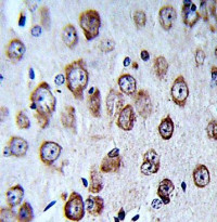
ARG55447 anti-PCSK9 antibody IHC-P image
Immunohistochemistry: Formalin-fixed and paraffin-embedded Human brain tissue stained with ARG55447 anti-PCSK9 antibody.
-
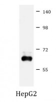
ARG55447 anti-PCSK9 antibody WB image
Western blot: 20 µg of HepG2 cell lysate stained with ARG55447 anti-PCSK9 antibody at 1:1000 dilution.
-
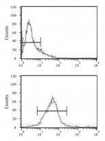
ARG55447 anti-PCSK9 antibody FACS image
Flow Cytometry: Jurkat cells stained with ARG55447 anti-PCSK9 antibody (bottom histogram) or without primary antibody control (top histogram), followed by incubation with FITC labelled secondary antibody.


