ARG55239
anti-PAX6 antibody
anti-PAX6 antibody for Flow cytometry,ICC/IF,IHC-Formalin-fixed paraffin-embedded sections,Western blot and Human
Controls and Markers antibody; Developmental Biology antibody; Gene Regulation antibody; Neuroscience antibody
Overview
| Product Description | Rabbit Polyclonal antibody recognizes PAX6 |
|---|---|
| Tested Reactivity | Hu |
| Predict Reactivity | Ms, Rat, Bov, Xenopus |
| Tested Application | FACS, ICC/IF, IHC-P, WB |
| Host | Rabbit |
| Clonality | Polyclonal |
| Isotype | IgG |
| Target Name | PAX6 |
| Antigen Species | Human |
| Immunogen | KLH-conjugated synthetic peptide corresponding to aa. 183-210 (Center) of Human PAX6. |
| Conjugation | Un-conjugated |
| Alternate Names | MGDA; Aniridia type II protein; AN; Oculorhombin; Paired box protein Pax-6; AN2; WAGR; FVH1; D11S812E |
Application Instructions
| Application Suggestion |
|
||||||||||
|---|---|---|---|---|---|---|---|---|---|---|---|
| Application Note | * The dilutions indicate recommended starting dilutions and the optimal dilutions or concentrations should be determined by the scientist. | ||||||||||
| Positive Control | U251 |
Properties
| Form | Liquid |
|---|---|
| Purification | Purification with Protein A and immunogen peptide. |
| Buffer | PBS and 0.09% (W/V) Sodium azide |
| Preservative | 0.09% (W/V) Sodium azide |
| Storage Instruction | For continuous use, store undiluted antibody at 2-8°C for up to a week. For long-term storage, aliquot and store at -20°C or below. Storage in frost free freezers is not recommended. Avoid repeated freeze/thaw cycles. Suggest spin the vial prior to opening. The antibody solution should be gently mixed before use. |
| Note | For laboratory research only, not for drug, diagnostic or other use. |
Bioinformation
| Database Links | |
|---|---|
| Gene Symbol | PAX6 |
| Gene Full Name | paired box 6 |
| Background | This gene encodes paired box gene 6, one of many human homologs of the Drosophila melanogaster gene prd. In addition to the hallmark feature of this gene family, a conserved paired box domain, the encoded protein also contains a homeo box domain. Both domains are known to bind DNA and function as regulators of gene transcription. This gene is expressed in the developing nervous system, and in developing eyes. Mutations in this gene are known to cause ocular disorders such as aniridia and Peter's anomaly. Alternatively spliced transcript variants encoding multiple isoforms have been observed for this gene. [provided by RefSeq, May 2012] |
| Function | Transcription factor with important functions in the development of the eye, nose, central nervous system and pancreas. Required for the differentiation of pancreatic islet alpha cells (By similarity). Competes with PAX4 in binding to a common element in the glucagon, insulin and somatostatin promoters. Regulates specification of the ventral neuron subtypes by establishing the correct progenitor domains (By similarity). Isoform 5a appears to function as a molecular switch that specifies target genes. [UniProt] |
| Cellular Localization | Nucleus. |
| Highlight | Related products: PAX6 antibodies; Anti-Rabbit IgG secondary antibodies; Related news: Choose the Best ZIKA Virus Antibodies Fight microcephaly with arigo |
| Research Area | Controls and Markers antibody; Developmental Biology antibody; Gene Regulation antibody; Neuroscience antibody |
| Calculated MW | 47 kDa |
| PTM | Ubiquitinated by TRIM11, leading to ubiquitination and proteasomal degradation. |
Images (4) Click the Picture to Zoom In
-

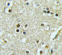
ARG55239 anti-PAX6 antibody ICC/IF image
Immunofluorescence: HeLa cells stained with ARG55239 anti-PAX6 antibody (green). Actin filaments have been labeled with Alexa Fluor 555 phalloidin (red).
-
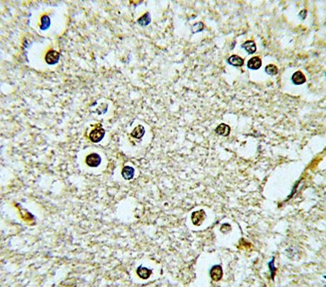
ARG55239 anti-PAX6 antibody IHC-P image
Immunohistochemistry: Formalin-fixed and paraffin-embedded Human brain tissue stained with ARG55239 anti-PAX6 antibody.
-

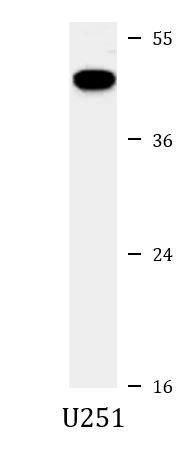
ARG55239 anti-PAX6 antibody WB image
Western blot: 35 µg of U251 cell lysate stained with ARG55239 anti-PAX6 antibody.
-
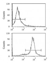
ARG55239 anti-PAX6 antibody FACS image
Flow Cytometry: HeLa cells stained with ARG55239 anti-PAX6 antibody (bottom histogram) or without primary antibody control (top histogram), followed by incubation with FITC labelled secondary antibody.