ARG40895
anti-PARP antibody
anti-PARP antibody for Flow cytometry,ICC/IF,IHC-Formalin-fixed paraffin-embedded sections,Western blot and Human,Mouse,Rat
Cancer antibody; Cell Biology and Cellular Response antibody; Cell Death antibody; Gene Regulation antibody; Metabolism antibody; Apoptosis Marker antibody; Mitochondria/Caspase Dependant Apoptosis Marker antibody

Overview
| Product Description | Rabbit Polyclonal antibody recognizes PARP |
|---|---|
| Tested Reactivity | Hu, Ms, Rat |
| Tested Application | FACS, ICC/IF, IHC-P, WB |
| Specificity | This antibody might reacts to full length and p25 fragment of PARP. |
| Host | Rabbit |
| Clonality | Polyclonal |
| Isotype | IgG |
| Target Name | PARP |
| Antigen Species | Human |
| Immunogen | Synthetic peptide derived from Human PARP. |
| Conjugation | Un-conjugated |
| Alternate Names | EC 2.4.2.30; Poly[ADP-ribose] synthase 1; PPOL; ADPRT; ARTD1; NAD; PARP-1; ADPRT 1; Poly [ADP-ribose] polymerase 1; PARP; ADP-ribosyltransferase diphtheria toxin-like 1; ADPRT1; pADPRT-1 |
Application Instructions
| Application Suggestion |
|
||||||||||
|---|---|---|---|---|---|---|---|---|---|---|---|
| Application Note | * The dilutions indicate recommended starting dilutions and the optimal dilutions or concentrations should be determined by the scientist. | ||||||||||
| Positive Control | Jurkat |
Properties
| Form | Liquid |
|---|---|
| Purification | Affinity purified. |
| Buffer | PBS (pH 7.4), 150 mM NaCl, 0.02% Sodium azide and 50% Glycerol. |
| Preservative | 0.02% Sodium azide |
| Stabilizer | 50% Glycerol |
| Storage Instruction | For continuous use, store undiluted antibody at 2-8°C for up to a week. For long-term storage, aliquot and store at -20°C. Storage in frost free freezers is not recommended. Avoid repeated freeze/thaw cycles. Suggest spin the vial prior to opening. The antibody solution should be gently mixed before use. |
| Note | For laboratory research only, not for drug, diagnostic or other use. |
Bioinformation
| Database Links | |
|---|---|
| Gene Symbol | PARP1 |
| Gene Full Name | poly (ADP-ribose) polymerase 1 |
| Background | This gene encodes a chromatin-associated enzyme, poly(ADP-ribosyl)transferase, which modifies various nuclear proteins by poly(ADP-ribosyl)ation. The modification is dependent on DNA and is involved in the regulation of various important cellular processes such as differentiation, proliferation, and tumor transformation and also in the regulation of the molecular events involved in the recovery of cell from DNA damage. In addition, this enzyme may be the site of mutation in Fanconi anemia, and may participate in the pathophysiology of type I diabetes. [provided by RefSeq, Jul 2008] |
| Function | Involved in the base excision repair (BER) pathway, by catalyzing the poly(ADP-ribosyl)ation of a limited number of acceptor proteins involved in chromatin architecture and in DNA metabolism. This modification follows DNA damages and appears as an obligatory step in a detection/signaling pathway leading to the reparation of DNA strand breaks. Mediates the poly(ADP-ribosyl)ation of APLF and CHFR. Positively regulates the transcription of MTUS1 and negatively regulates the transcription of MTUS2/TIP150. With EEF1A1 and TXK, forms a complex that acts as a T-helper 1 (Th1) cell-specific transcription factor and binds the promoter of IFN-gamma to directly regulate its transcription, and is thus involved importantly in Th1 cytokine production. Required for PARP9 and DTX3L recruitment to DNA damage sites. PARP1-dependent PARP9-DTX3L-mediated ubiquitination promotes the rapid and specific recruitment of 53BP1/TP53BP1, UIMC1/RAP80, and BRCA1 to DNA damage sites. [UniProt] |
| Cellular Localization | Nucleus. Nucleus, nucleolus. Note=Localizes at sites of DNA damage. [UniProt] |
| Research Area | Cancer antibody; Cell Biology and Cellular Response antibody; Cell Death antibody; Gene Regulation antibody; Metabolism antibody; Apoptosis Marker antibody; Mitochondria/Caspase Dependant Apoptosis Marker antibody |
| Calculated MW | 113 kDa |
| PTM | Phosphorylated by PRKDC and TXK. Poly-ADP-ribosylated by PARP2; poly-ADP-ribosylation mediates the recruitment of CHD1L to DNA damage sites (PubMed:19661379). ADP-ribosylated on serine by autocatalysis; serine ADP-ribosylation takes place following interaction with HPF1 (PubMed:28190768). S-nitrosylated, leading to inhibit transcription regulation activity. [UniProt] |
Images (4) Click the Picture to Zoom In
-
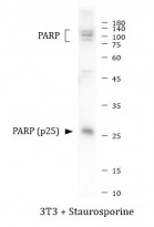
ARG40895 anti-PARP antibody WB image
Western blot: 3T3 cells were treated with Staurosporine. 20 μg of cell lysate stained with ARG40895 anti-PARP antibody at 1:1000 dilution.
-
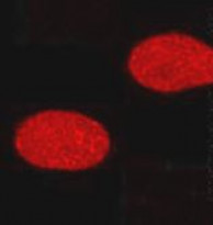
ARG40895 anti-PARP antibody ICC/IF image
Immunofluorescence: HeLa cells stained with ARG40895 anti-PARP antibody.
-
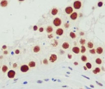
ARG40895 anti-PARP antibody IHC-P image
Immunohistochemistry: Paraffin-embedded Human testis tissue stained with ARG40895 anti-PARP antibody.
-
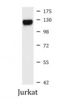
ARG40895 anti-PARP antibody WB image
Western blot: Jurkat cell lysate stained with ARG40895 anti-PARP antibody at 1:1000 dilution.


Customer's Feedback
Good

Review for anti-PARP antibody
Application:WB
Sample:3T3 + Staurosporine
Sample Loading Amount:20 μg
Primary Antibody Dilution Factor:1:1000
Primary Antibody Incubation Time:overnight
Primary Antibody Incubation Temperature:4 ºC


