ARG59016
anti-PARN antibody
anti-PARN antibody for Flow cytometry,ICC/IF,IHC-Formalin-fixed paraffin-embedded sections,Western blot and Human,Mouse,Rat
Overview
| Product Description | Rabbit Polyclonal antibody recognizes PARN |
|---|---|
| Tested Reactivity | Hu, Ms, Rat |
| Tested Application | FACS, ICC/IF, IHC-P, WB |
| Host | Rabbit |
| Clonality | Polyclonal |
| Isotype | IgG |
| Target Name | PARN |
| Antigen Species | Human |
| Immunogen | Recombinant protein corresponding to M1-Y301 of Human PARN. |
| Conjugation | Un-conjugated |
| Alternate Names | DAN; DKCB6; PFBMFT4; Poly(A)-specific ribonuclease PARN; EC 3.1.13.4; Deadenylating nuclease; Deadenylation nuclease; Polyadenylate-specific ribonuclease |
Application Instructions
| Application Suggestion |
|
||||||||||
|---|---|---|---|---|---|---|---|---|---|---|---|
| Application Note | IHC-P: Antigen Retrieval: Heat mediation was performed in Citrate buffer (pH 6.0) for 20 min. * The dilutions indicate recommended starting dilutions and the optimal dilutions or concentrations should be determined by the scientist. |
Properties
| Form | Liquid |
|---|---|
| Purification | Affinity purification with immunogen. |
| Buffer | 0.2% Na2HPO4, 0.9% NaCl, 0.05% Sodium azide and 4% Trehalose. |
| Preservative | 0.05% Sodium azide |
| Stabilizer | 4% Trehalose |
| Concentration | 0.5 mg/ml |
| Storage Instruction | For continuous use, store undiluted antibody at 2-8°C for up to a week. For long-term storage, aliquot and store at -20°C or below. Storage in frost free freezers is not recommended. Avoid repeated freeze/thaw cycles. Suggest spin the vial prior to opening. The antibody solution should be gently mixed before use. |
| Note | For laboratory research only, not for drug, diagnostic or other use. |
Bioinformation
| Database Links |
Swiss-port # O95453 Human Poly(A)-specific ribonuclease PARN Swiss-port # Q8VDG3 Mouse Poly(A)-specific ribonuclease PARN |
|---|---|
| Gene Symbol | PARN |
| Gene Full Name | poly(A)-specific ribonuclease |
| Background | The protein encoded by this gene is a 3'-exoribonuclease, with similarity to the RNase D family of 3'-exonucleases. It prefers poly(A) as the substrate, hence, efficiently degrades poly(A) tails of mRNAs. Exonucleolytic degradation of the poly(A) tail is often the first step in the decay of eukaryotic mRNAs. This protein is also involved in silencing of certain maternal mRNAs during oocyte maturation and early embryonic development, as well as in nonsense-mediated decay (NMD) of mRNAs that contain premature stop codons. Alternatively spliced transcript variants encoding different isoforms have been found for this gene. [provided by RefSeq, Aug 2008] |
| Function | 3'-exoribonuclease that has a preference for poly(A) tails of mRNAs, thereby efficiently degrading poly(A) tails. Exonucleolytic degradation of the poly(A) tail is often the first step in the decay of eukaryotic mRNAs and is also used to silence certain maternal mRNAs translationally during oocyte maturation and early embryonic development. Interacts with both the 3'-end poly(A) tail and the 5'-end cap structure during degradation, the interaction with the cap structure being required for an efficient degradation of poly(A) tails. Involved in nonsense-mediated mRNA decay, a critical process of selective degradation of mRNAs that contain premature stop codons. Also involved in degradation of inherently unstable mRNAs that contain AU-rich elements (AREs) in their 3'-UTR, possibly via its interaction with KHSRP. Probably mediates the removal of poly(A) tails of AREs mRNAs, which constitutes the first step of destabilization. [UniProt] |
| Cellular Localization | Nucleus. [UniProt] |
| Calculated MW | 73 kDa |
| PTM | Phosphorylation by MAPKAPK2, preventing GADD45A mRNA degradation after genotoxic stress. [UniProt] |
Images (6) Click the Picture to Zoom In
-
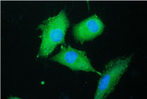
ARG59016 anti-PARN antibody ICC/IF image
Immunofluorescence: A549 cells were blocked with 10% goat serum and then stained with ARG59016 anti-PARN antibody (green) at 2 µg/ml dilution, overnight at 4°C. DAPI (blue) for nuclear staining.
-

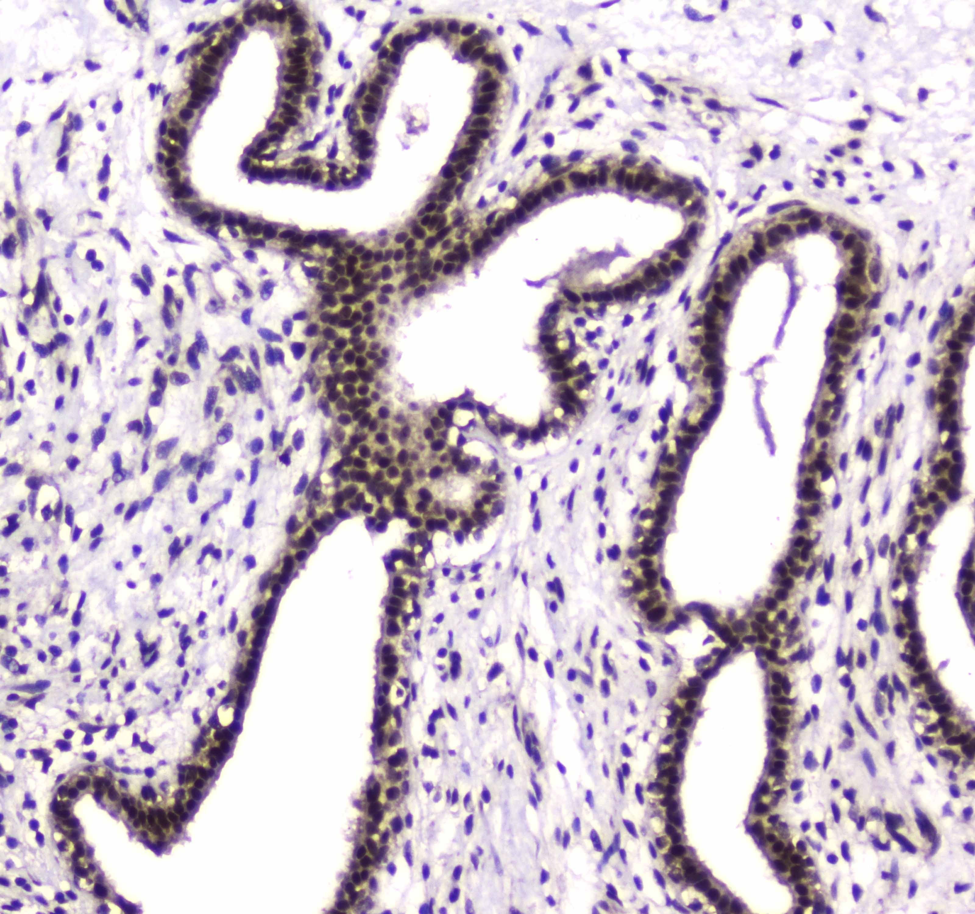
ARG59016 anti-PARN antibody IHC-P image
Immunohistochemistry: Paraffin-embedded Human mammary cancer tissue. Antigen Retrieval: Heat mediated was performed in Citrate buffer (pH 6.0, epitope retrieval solution) for 20 min. The tissue section was blocked with 10% goat serum. The tissue section was then stained with ARG59016 anti-PARN antibody at 1 µg/ml dilution, overnight at 4°C.
-

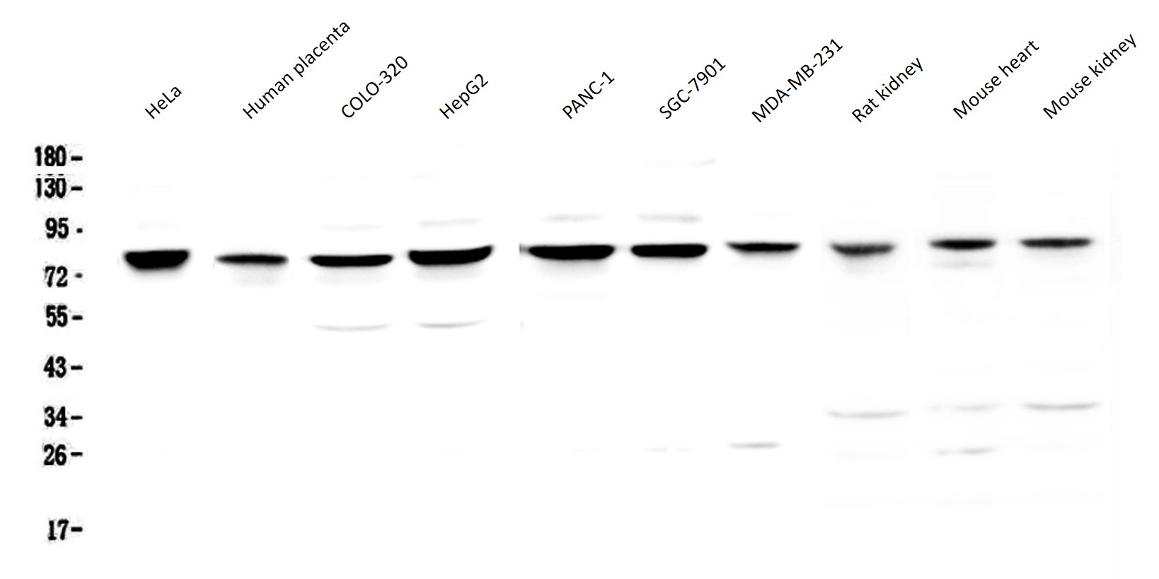
ARG59016 anti-PARN antibody WB image
Western blot: 50 µg of samples under reducing conditions. HeLa, Human placenta, COLO-320, HepG2, PANC-1, SGC-7901, MDA-MB-231, Rat kidney, Mouse heart and Mouse kidney lysates stained with ARG59016 anti-PARN antibody at 0.5 µg/ml, overnight at 4°C.
-
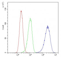
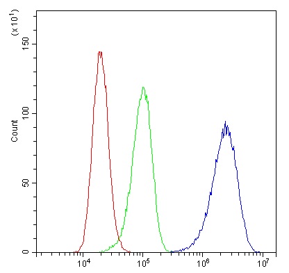
ARG59016 anti-PARN antibody FACS image
Flow Cytometry: A431 cells were blocked with 10% normal goat serum and then stained with ARG59016 anti-PARN antibody (blue) at 1 µg/10^6 cells for 30 min at 20°C, followed by incubation with DyLight®488 labelled secondary antibody. Isotype control antibody (green) was rabbit IgG (1 µg/10^6 cells) used under the same conditions. Unlabelled sample (red) was also used as a control.
-
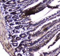
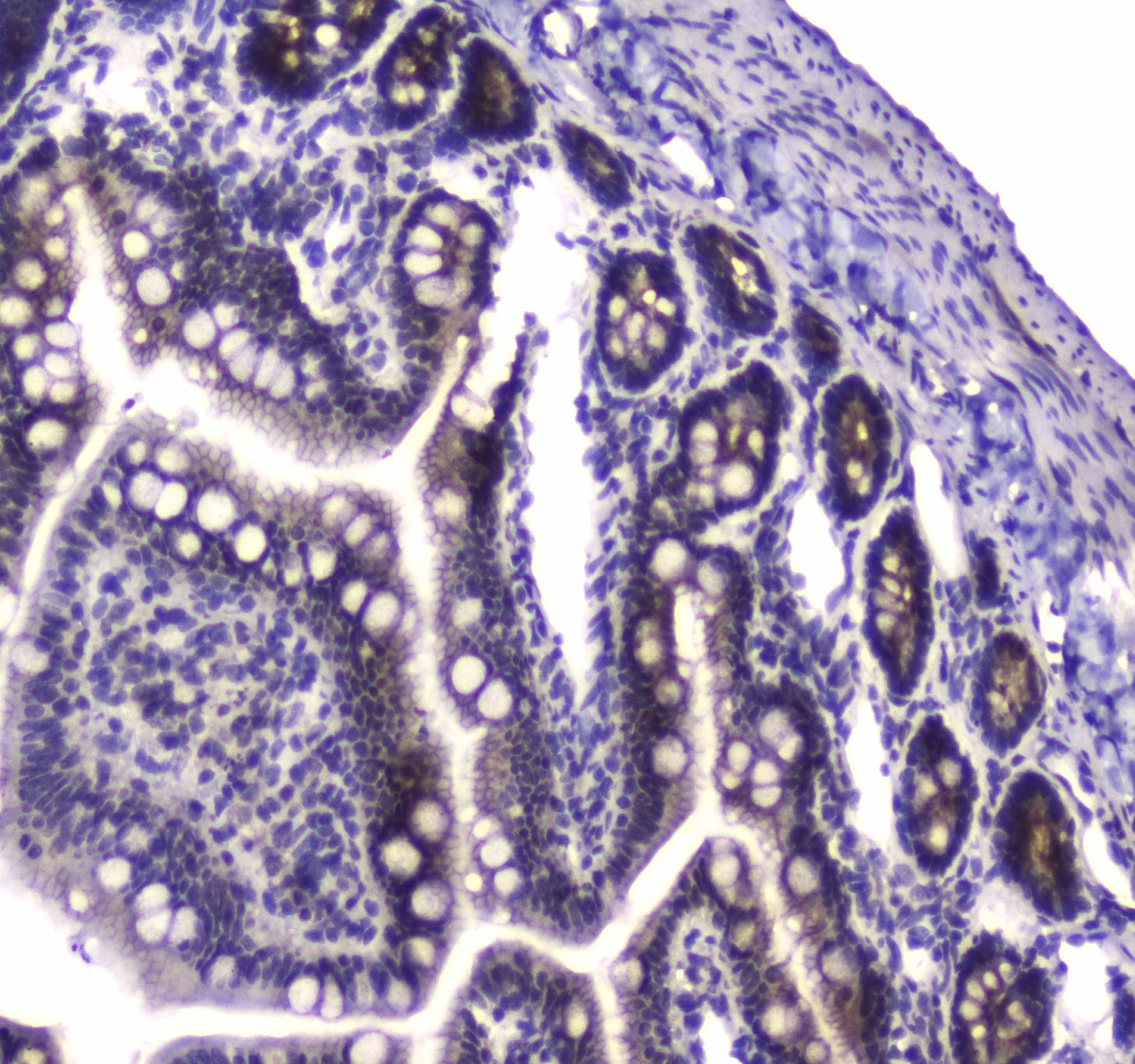
ARG59016 anti-PARN antibody IHC-P image
Immunohistochemistry: Paraffin-embedded Mouse small intestine tissue. Antigen Retrieval: Heat mediated was performed in Citrate buffer (pH 6.0, epitope retrieval solution) for 20 min. The tissue section was blocked with 10% goat serum. The tissue section was then stained with ARG59016 anti-PARN antibody at 1 µg/ml dilution, overnight at 4°C.
-
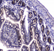
ARG59016 anti-PARN antibody IHC-P image
Immunohistochemistry: Paraffin-embedded Rat small intestine tissue. Antigen Retrieval: Heat mediated was performed in Citrate buffer (pH 6.0, epitope retrieval solution) for 20 min. The tissue section was blocked with 10% goat serum. The tissue section was then stained with ARG59016 anti-PARN antibody at 1 µg/ml dilution, overnight at 4°C.