ARG57133
anti-PARK7 / DJ1 antibody [1E12]
anti-PARK7 / DJ1 antibody [1E12] for Flow cytometry,Western blot and Human
Overview
| Product Description | Mouse Monoclonal antibody [1E12] recognizes PARK7 / DJ1 |
|---|---|
| Tested Reactivity | Hu |
| Tested Application | FACS, WB |
| Host | Mouse |
| Clonality | Monoclonal |
| Clone | 1E12 |
| Isotype | IgG2b, kappa |
| Target Name | PARK7 / DJ1 |
| Antigen Species | Human |
| Immunogen | Recombinant fragment around aa. 1-189 of Human PARK7 / DJ-1 |
| Conjugation | Un-conjugated |
| Alternate Names | DJ1; DJ-1; Oncogene DJ1; EC 3.5.1.-; Parkinson disease protein 7; HEL-S-67p; EC 3.1.2.-; Protein deglycase DJ-1 |
Application Instructions
| Application Suggestion |
|
||||||
|---|---|---|---|---|---|---|---|
| Application Note | * The dilutions indicate recommended starting dilutions and the optimal dilutions or concentrations should be determined by the scientist. |
Properties
| Form | Liquid |
|---|---|
| Purification | Purification with Protein A. |
| Buffer | PBS (pH 7.4), 0.02% Sodium azide and 10% Glycerol. |
| Preservative | 0.02% Sodium azide |
| Stabilizer | 10% Glycerol |
| Concentration | 1 mg/ml |
| Storage Instruction | For continuous use, store undiluted antibody at 2-8°C for up to a week. For long-term storage, aliquot and store at -20°C. Storage in frost free freezers is not recommended. Avoid repeated freeze/thaw cycles. Suggest spin the vial prior to opening. The antibody solution should be gently mixed before use. |
| Note | For laboratory research only, not for drug, diagnostic or other use. |
Bioinformation
| Database Links | |
|---|---|
| Gene Symbol | PARK7 |
| Gene Full Name | parkinson protein 7 |
| Background | The product of this gene belongs to the peptidase C56 family of proteins. It acts as a positive regulator of androgen receptor-dependent transcription. It may also function as a redox-sensitive chaperone, as a sensor for oxidative stress, and it apparently protects neurons against oxidative stress and cell death. Defects in this gene are the cause of autosomal recessive early-onset Parkinson disease 7. Two transcript variants encoding the same protein have been identified for this gene. [provided by RefSeq, Jul 2008] |
| Function | Protein deglycase that repairs methylglyoxal- and glyoxal-glycated amino acids and proteins, and releases repaired proteins and lactate or glycolate, respectively. Deglycates cysteines, arginines and lysines residues in proteins, and thus reactivates these proteins by reversing glycation by glyoxals. Acts on early glycation intermediates (hemithioacetals and aminocarbinols), preventing the formation of advanced glycation endproducts (AGE). Plays an important role in cell protection against oxidative stress and cell death acting as oxidative stress sensor and redox-sensitive chaperone and protease; functions probably related to its primary function. It is involved in neuroprotective mechanisms like the stabilization of NFE2L2 and PINK1 proteins, male fertility as a positive regulator of androgen signaling pathway as well as cell growth and transformation through, for instance, the modulation of NF-kappa-B signaling pathway. Its involvement in protein repair could also explain other unrelated functions. Eliminates hydrogen peroxide and protects cells against hydrogen peroxide-induced cell death. Required for correct mitochondrial morphology and function as well as for autophagy of dysfunctional mitochondria. Plays a role in regulating expression or stability of the mitochondrial uncoupling proteins SLC25A14 and SLC25A27 in dopaminergic neurons of the substantia nigra pars compacta and attenuates the oxidative stress induced by calcium entry into the neurons via L-type channels during pacemaking. Regulates astrocyte inflammatory responses, may modulate lipid rafts-dependent endocytosis in astrocytes and neuronal cells. Binds to a number of mRNAs containing multiple copies of GG or CC motifs and partially inhibits their translation but dissociates following oxidative stress. Metal-binding protein able to bind copper as well as toxic mercury ions, enhances the cell protection mechanism against induced metal toxicity. [UniProt] |
| Highlight | Related products: PARK7 antibodies; Anti-Mouse IgG secondary antibodies; Related news: Astrocyte-to-neuron conversion for Parkinson's disease treatment |
| Calculated MW | 20 kDa |
| PTM | Sumoylated on Lys-130 by PIAS2 or PIAS4; which is enhanced after ultraviolet irradiation and essential for cell-growth promoting activity and transforming activity. Cys-106 is easily oxidized to sulfinic acid. Undergoes cleavage of a C-terminal peptide and subsequent activation of protease activity in response to oxidative stress. |
Images (4) Click the Picture to Zoom In
-
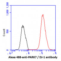
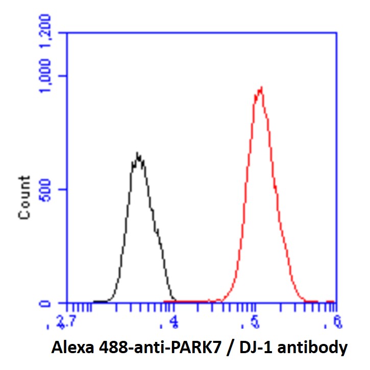
ARG57133 anti-PARK7 / DJ1 antibody [1E12] FACS image
Flow Cytometry: Hep3B cell line stained with ARG57133 anti-PARK7 / DJ1 antibody [1E12] at 2-5 µg for 1x10^6 cells (red line). Secondary antibody: Goat anti-Mouse IgG Alexa fluor 488 conjugate. Isotype control antibody: Mouse IgG (black line).
-
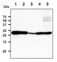
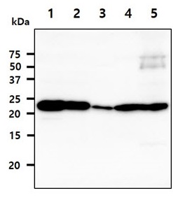
ARG57133 anti-PARK7 / DJ1 antibody [1E12] WB image
Western blot: 40 µg of 1) HeLa, 2) Jurkat, 3) NIH3T3, 4) Mouse brain, and 5) Mouse liver tissue lysate stained with ARG57133 anti-PARK7 / DJ1 antibody [1E12] at 1:1000.
-
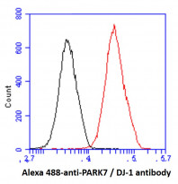
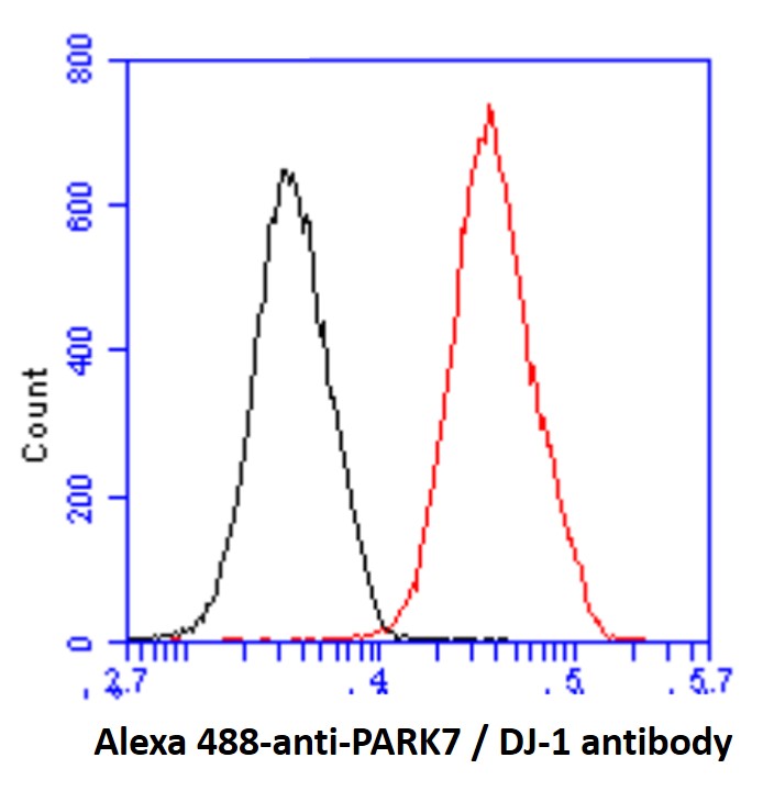
ARG57133 anti-PARK7 / DJ1 antibody [1E12] FACS image
Flow Cytometry: HeLa cell line stained with ARG57133 anti-PARK7 / DJ1 antibody [1E12] at 2-5 µg for 1x10^6 cells (red line). Secondary antibody: Goat anti-Mouse IgG Alexa fluor 488 conjugate. Isotype control antibody: Mouse IgG (black line).
-
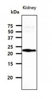
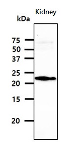
ARG57133 anti-PARK7 / DJ1 antibody [1E12] WB image
Western blot: 40 µg of kidney tissue lysate stained with ARG57133 anti-PARK7 / DJ1 antibody [1E12] at 1:1000.