ARG66841
anti-PAK5 / PAK7 antibody
anti-PAK5 / PAK7 antibody for IHC-Formalin-fixed paraffin-embedded sections,Western blot and Human,Mouse
Overview
| Product Description | Rabbit Polyclonal antibody recognizes PAK5 / PAK7 |
|---|---|
| Tested Reactivity | Hu, Ms |
| Predict Reactivity | Rat |
| Tested Application | IHC-P, WB |
| Host | Rabbit |
| Clonality | Polyclonal |
| Isotype | IgG |
| Target Name | PAK5 / PAK7 |
| Antigen Species | Human |
| Immunogen | Synthetic peptide between aa. 661-710 of Human PAK7. |
| Conjugation | Un-conjugated |
| Alternate Names | EC 2.7.11.1; Serine/threonine-protein kinase PAK 7; PAK5; p21-activated kinase 7; p21-activated kinase 5; PAK-7; PAK-5 |
Application Instructions
| Application Suggestion |
|
||||||
|---|---|---|---|---|---|---|---|
| Application Note | * The dilutions indicate recommended starting dilutions and the optimal dilutions or concentrations should be determined by the scientist. | ||||||
| Positive Control | Human brain and NIH/3T3 | ||||||
| Observed Size | 80 ~ 90 kDa |
Properties
| Form | Liquid |
|---|---|
| Purification | Affinity purification with immunogen. |
| Buffer | PBS, 0.02% Sodium azide, 50% Glycerol and 0.5% BSA. |
| Preservative | 0.02% Sodium azide |
| Stabilizer | 50% Glycerol and 0.5% BSA |
| Concentration | 1 mg/ml |
| Storage Instruction | For continuous use, store undiluted antibody at 2-8°C for up to a week. For long-term storage, aliquot and store at -20°C. Storage in frost free freezers is not recommended. Avoid repeated freeze/thaw cycles. Suggest spin the vial prior to opening. The antibody solution should be gently mixed before use. |
| Note | For laboratory research only, not for drug, diagnostic or other use. |
Bioinformation
| Database Links |
Swiss-port # Q8C015 Mouse Serine/threonine-protein kinase PAK 7 Swiss-port # Q9P286 Human Serine/threonine-protein kinase PAK 7 |
|---|---|
| Gene Symbol | PAK7 |
| Gene Full Name | p21 protein (Cdc42/Rac)-activated kinase 7 |
| Background | The protein encoded by this gene is a member of the PAK family of Ser/Thr protein kinases. PAK family members are known to be effectors of Rac/Cdc42 GTPases, which have been implicated in the regulation of cytoskeletal dynamics, proliferation, and cell survival signaling. This kinase contains a CDC42/Rac1 interactive binding (CRIB) motif, and has been shown to bind CDC42 in the presence of GTP. This kinase is predominantly expressed in brain. It is capable of promoting neurite outgrowth, and thus may play a role in neurite development. This kinase is associated with microtubule networks and induces microtubule stabilization. The subcellular localization of this kinase is tightly regulated during cell cycle progression. Alternatively spliced transcript variants encoding the same protein have been described. [provided by RefSeq, Jul 2008] |
| Function | Serine/threonine protein kinase that plays a role in a variety of different signaling pathways including cytoskeleton regulation, cell migration, proliferation or cell survival. Activation by various effectors including growth factor receptors or active CDC42 and RAC1 results in a conformational change and a subsequent autophosphorylation on several serine and/or threonine residues. Phosphorylates the proto-oncogene RAF1 and stimulates its kinase activity. Promotes cell survival by phosphorylating the BCL2 antagonist of cell death BAD. Phosphorylates CTNND1, probably to regulate cytoskeletal organization and cell morphology. Keeps microtubules stable through MARK2 inhibition and destabilizes the F-actin network leading to the disappearance of stress fibers and focal adhesions. [UniProt] |
| Cellular Localization | Mitochondrion. Cytoplasm. Nucleus. Note=Shuttles between the nucleus and the mitochondria, and mitochondrial localization is essential for the role in cell survival. [UniProt] |
| Calculated MW | 81 kDa |
| PTM | Autophosphorylated when activated by CDC42/p21. [UniProt] |
Images (3) Click the Picture to Zoom In
-
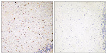
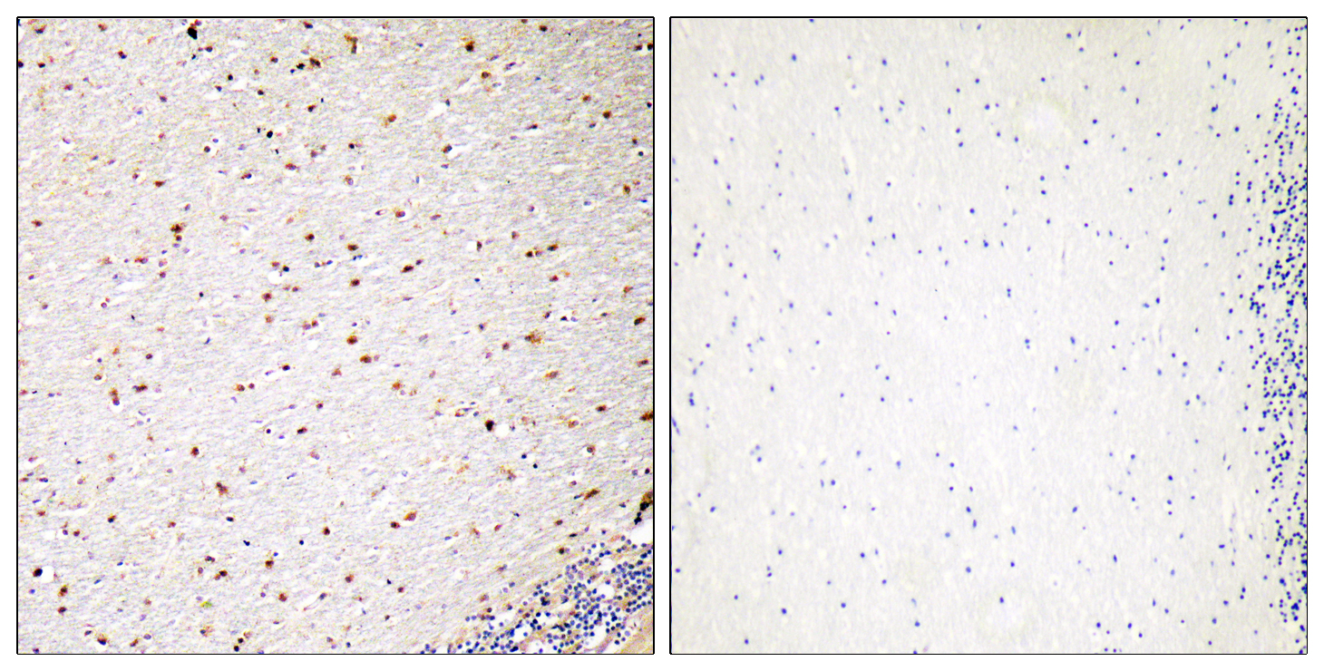
ARG66841 anti-PAK5 / PAK7 antibody IHC-P image
Immunohistochemistry: Paraffin-embedded Human brain tissue stained with ARG66841 anti-PAK5 / PAK7 antibody. The lane on the right is blocked with the peptide.
-
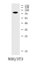
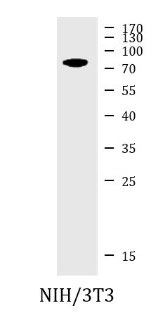
ARG66841 anti-PAK5 / PAK7 antibody WB image
Western blot: NIH/3T3 cell lysate stained with ARG66841 anti-PAK5 / PAK7 antibody.
-
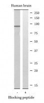
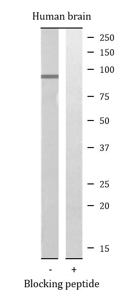
ARG66841 anti-PAK5 / PAK7 antibody WB image
Western blot: Human brain lysates stained with ARG66841 anti-PAK5 / PAK7 antibody. The lane on the right is blocked with the peptide.