ARG43115
anti-PAK4 antibody
anti-PAK4 antibody for ICC/IF,IHC-Formalin-fixed paraffin-embedded sections,Western blot and Human
Overview
| Product Description | Rabbit Polyclonal antibody recognizes PAK4 |
|---|---|
| Tested Reactivity | Hu |
| Tested Application | ICC/IF, IHC-P, WB |
| Host | Rabbit |
| Clonality | Polyclonal |
| Isotype | IgG |
| Target Name | PAK4 |
| Antigen Species | Human |
| Immunogen | A 13 amino acid peptide within aa. 210-260 of Human PAK4. |
| Conjugation | Un-conjugated |
| Alternate Names | EC 2.7.11.1; p21-activated kinase 4; Serine/threonine-protein kinase PAK 4; PAK-4 |
Application Instructions
| Application Suggestion |
|
||||||||
|---|---|---|---|---|---|---|---|---|---|
| Application Note | * The dilutions indicate recommended starting dilutions and the optimal dilutions or concentrations should be determined by the scientist. | ||||||||
| Positive Control | SW480 | ||||||||
| Observed Size | ~ 70 kDa |
Properties
| Form | Liquid |
|---|---|
| Purification | Affinity purification with immunogen. |
| Buffer | PBS and 0.02% Sodium azide. |
| Preservative | 0.02% Sodium azide |
| Concentration | 1 mg/ml |
| Storage Instruction | For continuous use, store undiluted antibody at 2-8°C for up to a week. For long-term storage, aliquot and store at -20°C or below. Storage in frost free freezers is not recommended. Avoid repeated freeze/thaw cycles. Suggest spin the vial prior to opening. The antibody solution should be gently mixed before use. |
| Note | For laboratory research only, not for drug, diagnostic or other use. |
Bioinformation
| Database Links |
Swiss-port # O96013 Human Serine/threonine-protein kinase PAK 4 |
|---|---|
| Gene Symbol | PAK4 |
| Gene Full Name | p21 protein (Cdc42/Rac)-activated kinase 4 |
| Background | PAK proteins, a family of serine/threonine p21-activating kinases, include PAK1, PAK2, PAK3 and PAK4. PAK proteins are critical effectors that link Rho GTPases to cytoskeleton reorganization and nuclear signaling. They serve as targets for the small GTP binding proteins Cdc42 and Rac and have been implicated in a wide range of biological activities. PAK4 interacts specifically with the GTP-bound form of Cdc42Hs and weakly activates the JNK family of MAP kinases. PAK4 is a mediator of filopodia formation and may play a role in the reorganization of the actin cytoskeleton. Multiple alternatively spliced transcript variants encoding distinct isoforms have been found for this gene. [provided by RefSeq, Jul 2008] |
| Function | Serine/threonine protein kinase that plays a role in a variety of different signaling pathways including cytoskeleton regulation, cell migration, growth, proliferation or cell survival. Activation by various effectors including growth factor receptors or active CDC42 and RAC1 results in a conformational change and a subsequent autophosphorylation on several serine and/or threonine residues. Phosphorylates and inactivates the protein phosphatase SSH1, leading to increased inhibitory phosphorylation of the actin binding/depolymerizing factor cofilin. Decreased cofilin activity may lead to stabilization of actin filaments. Phosphorylates LIMK1, a kinase that also inhibits the activity of cofilin. Phosphorylates integrin beta5/ITGB5 and thus regulates cell motility. Phosphorylates ARHGEF2 and activates the downstream target RHOA that plays a role in the regulation of assembly of focal adhesions and actin stress fibers. Stimulates cell survival by phosphorylating the BCL2 antagonist of cell death BAD. Alternatively, inhibits apoptosis by preventing caspase-8 binding to death domain receptors in a kinase independent manner. Plays a role in cell-cycle progression by controlling levels of the cell-cycle regulatory protein CDKN1A and by phosphorylating RAN. [UniProt] |
| Cellular Localization | Cytoplasm. Note=Seems to shuttle between cytoplasmic compartments depending on the activating effector. For example, can be found on the cell periphery after activation of growth-factor or integrin-mediated signaling pathways. [UniProt] |
| Calculated MW | 64 kDa |
| PTM | Autophosphorylated on serine residues when activated by CDC42/p21. Phosphorylated on tyrosine residues upon stimulation of FGFR2. [UniProt] |
Images (2) Click the Picture to Zoom In
-
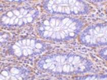
ARG43115 anti-PAK4 antibody IHC-P image
Immunohistochemistry: Paraffin-embedded Human colon tissue stained with ARG43115 anti-PAK4 antibody at 10 ug/ml dilution.
-
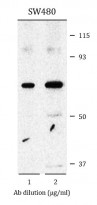
ARG43115 anti-PAK4 antibody WB image
Western blot: SW480 cell lysates stained with ARG43115 anti-PAK4 antibody at 1 µg/ml (left) and 2 µg/ml (right) dilution.


