ARG54657
anti-PAK2 antibody
anti-PAK2 antibody for ELISA,ICC/IF,IHC-Formalin-fixed paraffin-embedded sections,Western blot and Human,Mouse,Rat
Cancer antibody; Cell Biology and Cellular Response antibody; Cell Death antibody; Signaling Transduction antibody

Overview
| Product Description | Rabbit Polyclonal antibody recognizes PAK2 |
|---|---|
| Tested Reactivity | Hu, Ms, Rat |
| Tested Application | ELISA, ICC/IF, IHC-P, WB |
| Host | Rabbit |
| Clonality | Polyclonal |
| Isotype | IgG |
| Target Name | PAK2 |
| Immunogen | Synthetic peptide (14 aa) within aa. 20-70 of Human PAK2. |
| Conjugation | Un-conjugated |
| Alternate Names | Gamma-PAK; C-t-PAK2; PAK65; PAKgamma; S6/H4 kinase; p27; Serine/threonine-protein kinase PAK 2; p58; PAK-2; EC 2.7.11.1; p21-activated kinase 2; p34 |
Application Instructions
| Application Suggestion |
|
||||||||||
|---|---|---|---|---|---|---|---|---|---|---|---|
| Application Note | * The dilutions indicate recommended starting dilutions and the optimal dilutions or concentrations should be determined by the scientist. | ||||||||||
| Positive Control | Jurkat Cell Lysate |
Properties
| Form | Liquid |
|---|---|
| Purification | Affinity purification with immunogen. |
| Buffer | PBS and 0.02% Sodium azide |
| Preservative | 0.02% Sodium azide |
| Concentration | 1 mg/ml |
| Storage Instruction | For continuous use, store undiluted antibody at 2-8°C for up to a week. For long-term storage, aliquot and store at -20°C or below. Storage in frost free freezers is not recommended. Avoid repeated freeze/thaw cycles. Suggest spin the vial prior to opening. The antibody solution should be gently mixed before use. |
| Note | For laboratory research only, not for drug, diagnostic or other use. |
Bioinformation
| Database Links | |
|---|---|
| Gene Symbol | PAK2 |
| Gene Full Name | p21 protein (Cdc42/Rac)-activated kinase 2 |
| Background | PAK2 Antibody: The p21-activated kinases (PAKs) are serine-threonine kinases that bind to the active forms of Cdc42 and Rac. They are divided into two groups, the first of which include PAK1, 2 and 3, and can be activated by Cdc42/Rac binding. Group 1 PAKs contain an autoinhibitory domain whose activity is regulated by Cdc42/Rac binding. The group 1 PAKs are known to be involved in cellular processes such as gene transcription, apoptosis, and cell morphology and motility. Much less is known about the second group, which includes PAK4, 5 and 6, and are not activated by Cdc42/Rac binding. Of the six PAK proteins, only PAK2 is ubiquitously expressed and cleaved by caspase-3. This cleavage removes the amino-terminal regulatory domain and generates a constitutively active kinase fragment. Recent experiments have shown that following cleavage, the active fragment is myristoylated and directed to the plasma membrane and membrane ruffles where it promotes cell death via increased signaling through the c-Jun N-terminal kinase pathway, but without compromising mitochondrial integrity. | |
| Research Area | Cancer antibody; Cell Biology and Cellular Response antibody; Cell Death antibody; Signaling Transduction antibody |
| Calculated MW | 58 kDa |
| PTM | Full length PAK2 is autophosphorylated when activated by CDC42/p21. Following cleavage, both peptides, PAK-2p27 and PAK-2p34, become highly autophosphorylated, with PAK-2p27 being phosphorylated on serine and PAK-2p34 on threonine residues, respectively. Autophosphorylation of PAK-2p27 can occur in the absence of any effectors and is dependent on phosphorylation of Thr-402, because PAK-2p27 is acting as an exogenous substrate. During apoptosis proteolytically cleaved by caspase-3 or caspase-3-like proteases to yield active PAK-2p34. Ubiquitinated, leading to its proteasomal degradation. PAK-2p34 is myristoylated. |
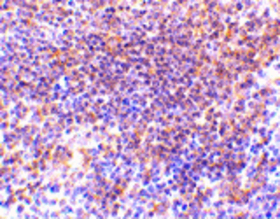
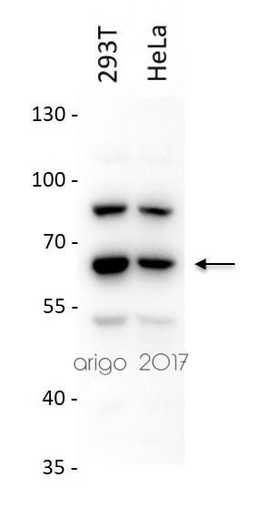
Images (3) Click the Picture to Zoom In
-
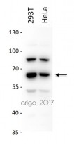
ARG54657 anti-PAK2 antibody WB image
Western blot: 20 µg of 293T and HeLa cell lysates stained with ARG54657 anti-PAK2 antibody at 2 µg/ml.
-
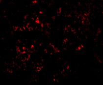
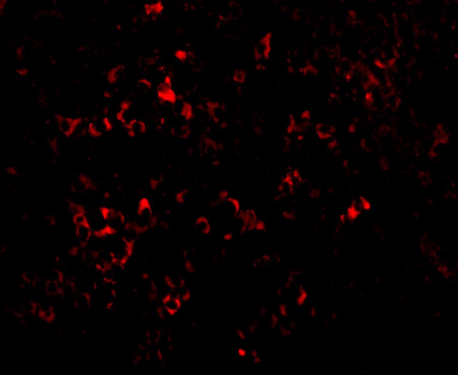
ARG54657 anti-PAK2 antibody ICC/IF image
Immunofluorescence: mouse spleen tissue stained with ARG54657 anti-PAK2 antibody at 20 μg/ml.
-
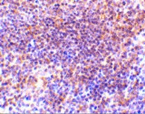
ARG54657 anti-PAK2 antibody IHC image
Immunohistochemistry: mouse spleen tissue stained with ARG54657 anti-PAK2 antibody at 10 μg/ml.

Customer's Feedback
Good
Review for anti-PAK2 antibody
Application:WB
Sample:293T and HeLa
Sample Loading Amount:20 µg
Primary Antibody Dilution Factor:2 µg/ml
Primary Antibody Incubation Time:overnight
Primary Antibody Incubation Temperature:4 ºC


