ARG40623
anti-PAK2 antibody
anti-PAK2 antibody for ICC/IF,IHC-Formalin-fixed paraffin-embedded sections,Western blot and Human,Monkey
Overview
| Product Description | Mouse Monoclonal antibody recognizes PAK2 |
|---|---|
| Tested Reactivity | Hu, Mk |
| Tested Application | ICC/IF, IHC-P, WB |
| Host | Mouse |
| Clonality | Monoclonal |
| Isotype | IgG1 |
| Target Name | PAK2 |
| Antigen Species | Human |
| Immunogen | Purified recombinant fragment of Human PAK2. |
| Conjugation | Un-conjugated |
| Alternate Names | Gamma-PAK; C-t-PAK2; PAK65; PAKgamma; S6/H4 kinase; p27; Serine/threonine-protein kinase PAK 2; p58; PAK-2; EC 2.7.11.1; p21-activated kinase 2; p34 |
Application Instructions
| Application Suggestion |
|
||||||||
|---|---|---|---|---|---|---|---|---|---|
| Application Note | * The dilutions indicate recommended starting dilutions and the optimal dilutions or concentrations should be determined by the scientist. |
Properties
| Form | Liquid |
|---|---|
| Purification | Unpurified |
| Buffer | Ascitic fluid and 0.03% Sodium azide. |
| Preservative | 0.03% Sodium azide |
| Storage Instruction | For continuous use, store undiluted antibody at 2-8°C for up to a week. For long-term storage, aliquot and store at -20°C or below. Storage in frost free freezers is not recommended. Avoid repeated freeze/thaw cycles. Suggest spin the vial prior to opening. The antibody solution should be gently mixed before use. |
| Note | For laboratory research only, not for drug, diagnostic or other use. |
Bioinformation
| Database Links |
Swiss-port # Q13177 Human Serine/threonine-protein kinase PAK 2 |
|---|---|
| Gene Symbol | PAK2 |
| Gene Full Name | p21 protein (Cdc42/Rac)-activated kinase 2 |
| Background | The p21 activated kinases (PAK) are critical effectors that link Rho GTPases to cytoskeleton reorganization and nuclear signaling. The PAK proteins are a family of serine/threonine kinases that serve as targets for the small GTP binding proteins, CDC42 and RAC1, and have been implicated in a wide range of biological activities. The protein encoded by this gene is activated by proteolytic cleavage during caspase-mediated apoptosis, and may play a role in regulating the apoptotic events in the dying cell. [provided by RefSeq, Jul 2008] |
| Function | Serine/threonine protein kinase that plays a role in a variety of different signaling pathways including cytoskeleton regulation, cell motility, cell cycle progression, apoptosis or proliferation. Acts as downstream effector of the small GTPases CDC42 and RAC1. Activation by the binding of active CDC42 and RAC1 results in a conformational change and a subsequent autophosphorylation on several serine and/or threonine residues. Full-length PAK2 stimulates cell survival and cell growth. Phosphorylates MAPK4 and MAPK6 and activates the downstream target MAPKAPK5, a regulator of F-actin polymerization and cell migration. Phosphorylates JUN and plays an important role in EGF-induced cell proliferation. Phosphorylates many other substrates including histone H4 to promote assembly of H3.3 and H4 into nucleosomes, BAD, ribosomal protein S6, or MBP. Additionally, associates with ARHGEF7 and GIT1 to perform kinase-independent functions such as spindle orientation control during mitosis. On the other hand, apoptotic stimuli such as DNA damage lead to caspase-mediated cleavage of PAK2, generating PAK-2p34, an active p34 fragment that translocates to the nucleus and promotes cellular apoptosis involving the JNK signaling pathway. Caspase-activated PAK2 phosphorylates MKNK1 and reduces cellular translation. [UniProt] |
| Cellular Localization | Serine/threonine-protein kinase PAK 2: Cytoplasm. Note=MYO18A mediates the cellular distribution of the PAK2-ARHGEF7-GIT1 complex to the inner surface of the cell membrane. PAK-2p34: Nucleus. Cytoplasm, perinuclear region. Membrane; Lipid-anchor. Note=Interaction with ARHGAP10 probably changes PAK-2p34 location to cytoplasmic perinuclear region. Myristoylation changes PAK-2p34 location to the membrane. [UniProt] |
| Calculated MW | 58 kDa |
| PTM | Full length PAK2 is autophosphorylated when activated by CDC42/p21. Following cleavage, both peptides, PAK-2p27 and PAK-2p34, become highly autophosphorylated, with PAK-2p27 being phosphorylated on serine and PAK-2p34 on threonine residues, respectively. Autophosphorylation of PAK-2p27 can occur in the absence of any effectors and is dependent on phosphorylation of Thr-402, because PAK-2p27 is acting as an exogenous substrate. During apoptosis proteolytically cleaved by caspase-3 or caspase-3-like proteases to yield active PAK-2p34. Ubiquitinated, leading to its proteasomal degradation. PAK-2p34 is myristoylated. [UniProt] |
Images (3) Click the Picture to Zoom In
-

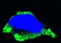
ARG40623 anti-PAK2 antibody ICC/IF image
Immunofluorescence: HeLa cells stained with ARG40623 anti-PAK2 antibody (green).
Blue: DRAQ5 fluorescent DNA dye.
-
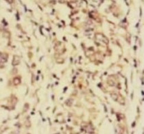
ARG40623 anti-PAK2 antibody IHC-P image
Immunohistochemistry: Paraffin-embedded Human gastric cancer tissue stained with ARG40623 anti-PAK2 antibody.
-

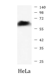
ARG40623 anti-PAK2 antibody WB image
Western blot: HeLa cell lysate stained with ARG40623 anti-PAK2 antibody at 1:5000 dilution.