ARG54868
anti-PACSIN2 antibody
anti-PACSIN2 antibody for ICC/IF,IHC-Formalin-fixed paraffin-embedded sections,Western blot and Human,Mouse
Signaling Transduction antibody
Overview
| Product Description | Rabbit Polyclonal antibody recognizes PACSIN2 |
|---|---|
| Tested Reactivity | Hu, Ms |
| Tested Application | ICC/IF, IHC-P, WB |
| Host | Rabbit |
| Clonality | Polyclonal |
| Isotype | IgG |
| Target Name | PACSIN2 |
| Antigen Species | Human |
| Immunogen | KLH-conjugated synthetic peptide corresponding to aa. 342-371 (C-terminus) of Human PACSIN2. |
| Conjugation | Un-conjugated |
| Alternate Names | SDPII; Protein kinase C and casein kinase substrate in neurons protein 2; Syndapin-II; Syndapin-2 |
Application Instructions
| Application Suggestion |
|
||||||||
|---|---|---|---|---|---|---|---|---|---|
| Application Note | * The dilutions indicate recommended starting dilutions and the optimal dilutions or concentrations should be determined by the scientist. | ||||||||
| Positive Control | Daudi |
Properties
| Form | Liquid |
|---|---|
| Purification | Purification with Protein G. |
| Buffer | PBS and 0.09% (W/V) Sodium azide |
| Preservative | 0.09% (W/V) Sodium azide |
| Storage Instruction | For continuous use, store undiluted antibody at 2-8°C for up to a week. For long-term storage, aliquot and store at -20°C or below. Storage in frost free freezers is not recommended. Avoid repeated freeze/thaw cycles. Suggest spin the vial prior to opening. The antibody solution should be gently mixed before use. |
| Note | For laboratory research only, not for drug, diagnostic or other use. |
Bioinformation
| Database Links |
Swiss-port # Q9UNF0 Human Protein kinase C and casein kinase substrate in neurons protein 2 Swiss-port # Q9WVE8 Mouse Protein kinase C and casein kinase substrate in neurons protein 2 |
|---|---|
| Gene Symbol | PACSIN2 |
| Gene Full Name | protein kinase C and casein kinase substrate in neurons 2 |
| Background | This gene is a member of the protein kinase C and casein kinase substrate in neurons family. The encoded protein is involved in linking the actin cytoskeleton with vesicle formation by regulating tubulin polymerization. Alternative splicing results in multiple transcript variants. [provided by RefSeq, May 2010] |
| Function | Lipid-binding protein that is able to promote the tubulation of the phosphatidic acid-containing membranes it preferentially binds. Plays a role in intracellular vesicle-mediated transport. Involved in the endocytosis of cell-surface receptors like the EGF receptor, contributing to its internalization in the absence of EGF stimulus. May also play a role in the formation of caveolae at the cell membrane. Recruits DNM2 to caveolae, and thereby plays a role in caveola-mediated endocytosis. [UniProt] |
| Cellular Localization | Cytoplasm. Cytoplasm, cytoskeleton. Cytoplasmic vesicle membrane; Peripheral membrane protein; Cytoplasmic side. Early endosome Recycling endosome membrane. Cell projection, ruffle membrane; Peripheral membrane protein; Cytoplasmic side. Cell membrane; Peripheral membrane protein; Cytoplasmic side. Cell projection. Membrane, caveola. Note=Detected at the neck of flask-shaped caveolae. Localization to tubular recycling endosomes probably requires interaction with MICALL1 and EHD1 |
| Research Area | Signaling Transduction antibody |
| Calculated MW | 56 kDa |
| PTM | Phosphorylated by casein kinase 2 (CK2) and protein kinase C (PKC). |
Images (3) Click the Picture to Zoom In
-
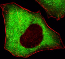
ARG54868 anti-PACSIN2 antibody ICC/IF image
Immunofluorescence: HeLa cells stained with ARG54868 anti-PACSIN2 antibody (green) at 1:100 dilution. Cytoplasmic actin was counterstained with Dylight Fluor® 554 conjugated Phalloidin (red).
-
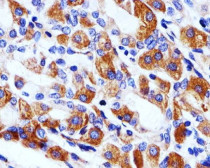
ARG54868 anti-PACSIN2 antibody IHC-P image
Immunohistochemistry: Paraffin-embedded Human stomach tissue stained with ARG54868 anti-PACSIN2 antibody at 1:100 dilution.
-
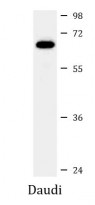
ARG54868 anti-PACSIN2 antibody WB image
Western blot: 35 µg of Daudi cell lysate stained with ARG54868 anti-PACSIN2 antibody.


