ARG63613
anti-PACSIN1 antibody
anti-PACSIN1 antibody for IHC-Formalin-fixed paraffin-embedded sections,Western blot and Human
Signaling Transduction antibody
Overview
| Product Description | Goat Polyclonal antibody recognizes PACSIN1 |
|---|---|
| Tested Reactivity | Hu |
| Predict Reactivity | Cow, Dog, Pig |
| Tested Application | IHC-P, WB |
| Specificity | Reported variants represent identical protein (NP_065855.1; NP_001186512.1). |
| Host | Goat |
| Clonality | Polyclonal |
| Isotype | IgG |
| Target Name | PACSIN1 |
| Antigen Species | Human |
| Immunogen | SSSYDEASLAPEET-C |
| Conjugation | Un-conjugated |
| Alternate Names | SDPI; Protein kinase C and casein kinase substrate in neurons protein 1; Syndapin-1 |
Application Instructions
| Application Suggestion |
|
||||||
|---|---|---|---|---|---|---|---|
| Application Note | WB: Recommend incubate at RT for 1h. IHC-P: Antigen Retrieval: Steam tissue section in Citrate buffer (pH 6.0). * The dilutions indicate recommended starting dilutions and the optimal dilutions or concentrations should be determined by the scientist. |
Properties
| Form | Liquid |
|---|---|
| Purification | Purified from goat serum by antigen affinity chromatography. |
| Buffer | Tris saline (pH 7.3), 0.02% Sodium azide and 0.5% BSA. |
| Preservative | 0.02% Sodium azide |
| Stabilizer | 0.5% BSA |
| Concentration | 0.5 mg/ml |
| Storage Instruction | For continuous use, store undiluted antibody at 2-8°C for up to a week. For long-term storage, aliquot and store at -20°C or below. Storage in frost free freezers is not recommended. Avoid repeated freeze/thaw cycles. Suggest spin the vial prior to opening. The antibody solution should be gently mixed before use. |
| Note | For laboratory research only, not for drug, diagnostic or other use. |
Bioinformation
| Database Links |
Swiss-port # Q9BY11 Human Protein kinase C and casein kinase substrate in neurons protein 1 |
|---|---|
| Gene Symbol | PACSIN1 |
| Gene Full Name | protein kinase C and casein kinase substrate in neurons 1 |
| Function | Plays a role in the reorganization of the microtubule cytoskeleton via its interaction with MAPT; this decreases microtubule stability and inhibits MAPT-induced microtubule polymerization. Plays a role in cellular transport processes by recruiting DNM1, DNM2 and DNM3 to membranes. Plays a role in the reorganization of the actin cytoskeleton and in neuron morphogenesis via its interaction with COBL and WASL, and by recruiting COBL to the cell cortex. Plays a role in the regulation of neurite formation, neurite branching and the regulation of neurite length. Required for normal synaptic vesicle endocytosis; this process retrieves previously released neurotransmitters to accommodate multiple cycles of neurotransmission. Required for normal excitatory and inhibitory synaptic transmission (By similarity). Binds to membranes via its F-BAR domain and mediates membrane tubulation. [UniProt] |
| Research Area | Signaling Transduction antibody |
| Calculated MW | 51 kDa |
| PTM | Phosphorylated by casein kinase 2 (CK2) and protein kinase C (PKC). |
Images (3) Click the Picture to Zoom In
-
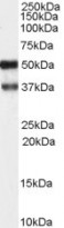
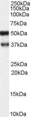
ARG63613 anti-PACSIN1 antibody WB image
Western Blot: Human Brain (hippocampus) lysate (35 µg protein in RIPA buffer) stained with ARG63613 anti-PACSIN1 antibody at 1 µg/ml dilution.
-
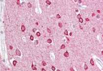
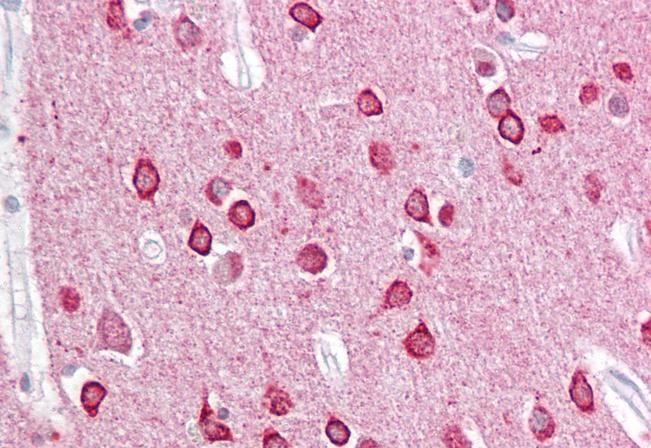
ARG63613 anti-PACSIN1 antibody IHC-P image
Immunohistochemistry: Paraffin-embedded Human cortex tissue. Antigen Retrieval: Steam tissue section in Citrate buffer (pH 6.0). The tissue section was stained with ARG63613 anti-PACSIN1 antibody at 2.5 µg/ml dilution followed by AP-staining.
-
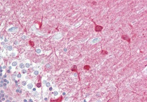
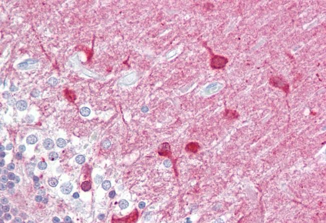
ARG63613 anti-PACSIN1 antibody IHC-P image
Immunohistochemistry: Paraffin-embedded Human cerebellum tissue. Antigen Retrieval: Steam tissue section in Citrate buffer (pH 6.0). The tissue section was stained with ARG63613 anti-PACSIN1 antibody at 2.5 µg/ml dilution followed by AP-staining.