ARG42588
anti-Optineurin antibody
anti-Optineurin antibody for Flow cytometry,IHC-Formalin-fixed paraffin-embedded sections,Western blot and Human,Mouse,Rat
Overview
| Product Description | Rabbit Polyclonal antibody recognizes Optineurin |
|---|---|
| Tested Reactivity | Hu, Ms, Rat |
| Tested Application | FACS, IHC-P, WB |
| Host | Rabbit |
| Clonality | Polyclonal |
| Isotype | IgG |
| Target Name | Optineurin |
| Antigen Species | Human |
| Immunogen | Recombinant protein corresponding to R241-I577 of Human Optineurin. |
| Conjugation | Un-conjugated |
| Alternate Names | GLC1E; Transcription factor IIIA-interacting protein; E3-14.7K-interacting protein; Optineurin; NEMO-related protein; Huntingtin-interacting protein 7; TFIIIA-INTP; Optic neuropathy-inducing protein; HYPL; Huntingtin-interacting protein L; FIP2; ALS12; Huntingtin yeast partner L; HIP7; NRP; FIP-2; HIP-7; TFIIIA-IntP |
Application Instructions
| Application Suggestion |
|
||||||||
|---|---|---|---|---|---|---|---|---|---|
| Application Note | * The dilutions indicate recommended starting dilutions and the optimal dilutions or concentrations should be determined by the scientist. | ||||||||
| Observed Size | ~ 66 kDa |
Properties
| Form | Liquid |
|---|---|
| Purification | Affinity purification with immunogen. |
| Buffer | 0.2% Na2HPO4, 0.9% NaCl, 0.05% Sodium azide and 5% BSA. |
| Preservative | 0.05% Sodium azide |
| Stabilizer | 5% BSA |
| Concentration | 0.5 mg/ml |
| Storage Instruction | For continuous use, store undiluted antibody at 2-8°C for up to a week. For long-term storage, aliquot and store at -20°C or below. Storage in frost free freezers is not recommended. Avoid repeated freeze/thaw cycles. Suggest spin the vial prior to opening. The antibody solution should be gently mixed before use. |
| Note | For laboratory research only, not for drug, diagnostic or other use. |
Bioinformation
| Database Links | |
|---|---|
| Gene Symbol | OPTN |
| Gene Full Name | optineurin |
| Background | This gene encodes the coiled-coil containing protein optineurin. Optineurin may play a role in normal-tension glaucoma and adult-onset primary open angle glaucoma. Optineurin interacts with adenovirus E3-14.7K protein and may utilize tumor necrosis factor-alpha or Fas-ligand pathways to mediate apoptosis, inflammation or vasoconstriction. Optineurin may also function in cellular morphogenesis and membrane trafficking, vesicle trafficking, and transcription activation through its interactions with the RAB8, huntingtin, and transcription factor IIIA proteins. Alternative splicing results in multiple transcript variants encoding the same protein. [provided by RefSeq, Jul 2008] |
| Function | Plays an important role in the maintenance of the Golgi complex, in membrane trafficking, in exocytosis, through its interaction with myosin VI and Rab8 (PubMed:27534431). Links myosin VI to the Golgi complex and plays an important role in Golgi ribbon formation (PubMed:27534431). Plays a role in the activation of innate immune response during viral infection. Mechanistically, recruits TBK1 at the Golgi apparatus, promoting its trans-phosphorylation after RLR or TLR3 stimulation (PubMed:27538435). In turn, activated TBK1 phosphorylates its downstream partner IRF3 to produce IFN-beta. Plays a neuroprotective role in the eye and optic nerve. May act by regulating membrane trafficking and cellular morphogenesis via a complex that contains Rab8 and hungtingtin (HD). Mediates the interaction of Rab8 with the probable GTPase-activating protein TBC1D17 during Rab8-mediated endocytic trafficking, such as of transferrin receptor (TFRC/TfR); regulates Rab8 recruitment to tubules emanating from the endocytic recycling compartment. Autophagy receptor that interacts directly with both the cargo to become degraded and an autophagy modifier of the MAP1 LC3 family; targets ubiquitin-coated bacteria (xenophagy), such as cytoplasmic Salmonella enterica, and appears to function in the same pathway as SQSTM1 and CALCOCO2/NDP52. (Microbial infection) May constitute a cellular target for adenovirus E3 14.7 and Bluetongue virus protein NS3 to inhibit innate immune response. [UniProt] |
| Cellular Localization | Cytoplasm, perinuclear region. Golgi apparatus. Golgi apparatus, trans-Golgi network. Cytoplasmic vesicle, autophagosome. Cytoplasmic vesicle. Recycling endosome. Note=Found in the perinuclear region and associates with the Golgi apparatus (PubMed:27534431). Colocalizes with MYO6 and RAB8 at the Golgi complex and in vesicular structures close to the plasma membrane. Localizes to LC3-positive cytoplasmic vesicles upon induction of autophagy. [UniProt] |
| Calculated MW | 66 kDa |
| PTM | Phosphorylated by TBK1, leading to restrict bacterial proliferation in case of infection. Phosphorylation is induced by phorbol esters and decreases its half-time. [UniProt] |
Images (7) Click the Picture to Zoom In
-

ARG42588 anti-Optineurin antibody IHC-P image
Immunohistochemistry: Human gall bladder adenosquamous carcinoma stained with ARG42588 anti-Optineurin antibody at 2 μg/ml dilution.
-

ARG42588 anti-Optineurin antibody WB image
Western blot: Hela, HT1080 and HepG2 stained with ARG42588 anti-Optineurin antibody at 0.5 μg/mL dilution.
-
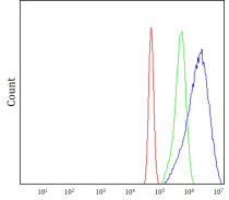
ARG42588 anti-Optineurin antibody FACS image
Flow Cytometry: A431 stained with ARG42588 anti-Optineurin antibody at 1 μg/1x10^6 cells dilution.
-

ARG42588 anti-Optineurin antibody IHC-P image
Immunohistochemistry: Rat brain stained with ARG42588 anti-Optineurin antibody at 2 μg/ml dilution.
-

ARG42588 anti-Optineurin antibody WB image
Western blot: Rat heart, Rat brain stained with ARG42588 anti-Optineurin antibody at 0.5 μg/mL dilution.
-

ARG42588 anti-Optineurin antibody IHC-P image
Immunohistochemistry: Mouse brain stained with ARG42588 anti-Optineurin antibody at 2 μg/ml dilution.
-

ARG42588 anti-Optineurin antibody WB image
Western blot: Mouse heart, Mouse brain stained with ARG42588 anti-Optineurin antibody at 0.5 μg/mL dilution.


