ARG57151
anti-OTUB1 antibody [17E10]
anti-OTUB1 antibody [17E10] for Western blot and Human
Overview
| Product Description | Mouse Monoclonal antibody [17E10] recognizes OTUB1 |
|---|---|
| Tested Reactivity | Hu |
| Tested Application | WB |
| Host | Mouse |
| Clonality | Monoclonal |
| Clone | 17E10 |
| Isotype | IgG1, kappa |
| Target Name | OTUB1 |
| Antigen Species | Human |
| Immunogen | Recombinant fragment around aa. 1-271 of Human OTUB1 |
| Conjugation | Un-conjugated |
| Alternate Names | HSPC263; hOTU1; Ubiquitin-specific-processing protease OTUB1; Deubiquitinating enzyme OTUB1; OTB1; OTU1; Otubain-1; OTU domain-containing ubiquitin aldehyde-binding protein 1; Ubiquitin thioesterase OTUB1; EC 3.4.19.12 |
Application Instructions
| Application Suggestion |
|
||||
|---|---|---|---|---|---|
| Application Note | * The dilutions indicate recommended starting dilutions and the optimal dilutions or concentrations should be determined by the scientist. |
Properties
| Form | Liquid |
|---|---|
| Purification | Purification with Protein A. |
| Buffer | PBS (pH 7.4), 0.02% Sodium azide and 10% Glycerol. |
| Preservative | 0.02% Sodium azide |
| Stabilizer | 10% Glycerol |
| Concentration | 1 mg/ml |
| Storage Instruction | For continuous use, store undiluted antibody at 2-8°C for up to a week. For long-term storage, aliquot and store at -20°C. Storage in frost free freezers is not recommended. Avoid repeated freeze/thaw cycles. Suggest spin the vial prior to opening. The antibody solution should be gently mixed before use. |
| Note | For laboratory research only, not for drug, diagnostic or other use. |
Bioinformation
| Database Links | |
|---|---|
| Gene Symbol | OTUB1 |
| Gene Full Name | OTU deubiquitinase, ubiquitin aldehyde binding 1 |
| Background | The product of this gene is a member of the OTU (ovarian tumor) superfamily of predicted cysteine proteases. The encoded protein is a highly specific ubiquitin iso-peptidase, and cleaves ubiquitin from branched poly-ubiquitin chains but not from ubiquitinated substrates. It interacts with another ubiquitin protease and an E3 ubiquitin ligase that inhibits cytokine gene transcription in the immune system. It is proposed to function in specific ubiquitin-dependent pathways, possibly by providing an editing function for polyubiquitin chain growth. Alternative splicing results in multiple transcript variants. [provided by RefSeq, Jul 2008] |
| Function | Hydrolase that can specifically remove 'Lys-48'-linked conjugated ubiquitin from proteins and plays an important regulatory role at the level of protein turnover by preventing degradation. Regulator of T-cell anergy, a phenomenon that occurs when T-cells are rendered unresponsive to antigen rechallenge and no longer respond to their cognate antigen. Acts via its interaction with RNF128/GRAIL, a crucial inductor of CD4 T-cell anergy. Isoform 1 destabilizes RNF128, leading to prevent anergy. In contrast, isoform 2 stabilizes RNF128 and promotes anergy. Surprisingly, it regulates RNF128-mediated ubiquitination, but does not deubiquitinate polyubiquitinated RNF128. Deubiquitinates estrogen receptor alpha (ESR1). Mediates deubiquitination of 'Lys-48'-linked polyubiquitin chains, but not 'Lys-63'-linked polyubiquitin chains. Not able to cleave di-ubiquitin. Also capable of removing NEDD8 from NEDD8 conjugates, but with a much lower preference compared to 'Lys-48'-linked ubiquitin. Plays a key non-catalytic role in DNA repair regulation by inhibiting activity of RNF168, an E3 ubiquitin-protein ligase that promotes accumulation of 'Lys-63'-linked histone H2A and H2AX at DNA damage sites. Inhibits RNF168 independently of ubiquitin thioesterase activity by binding and inhibiting UBE2N/UBC13, the E2 partner of RNF168, thereby limiting spreading of 'Lys-63'-linked histone H2A and H2AX marks. Inhibition occurs by binding to free ubiquitin: free ubiquitin acts as an allosteric regulator that increases affinity for UBE2N/UBC13 and disrupts interaction with UBE2V1. The OTUB1-UBE2N/UBC13-free ubiquitin complex adopts a configuration that mimics a cleaved 'Lys48'-linked di-ubiquitin chain. [UniProt] |
| Calculated MW | 31 kDa |
Images (1) Click the Picture to Zoom In
-
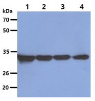
ARG57151 anti-OTUB1 antibody [17E10] WB image
Western blot: 40 µg of 1) A549, 2) HeLa, 3) HepG2, and 4) 293T cell lysates stained with ARG57151 anti-OTUB1 antibody [17E10] at 1:5000.


